
Fig. 1
- ID
- ZDB-IMAGE-130320-1
- Antibodies
- Publication
- Sainath et al., 2013 - Plexin a3 and turnout regulate motor axonal branch morphogenesis in zebrafish
- All Figures
- Figures for Sainath et al., 2013
|
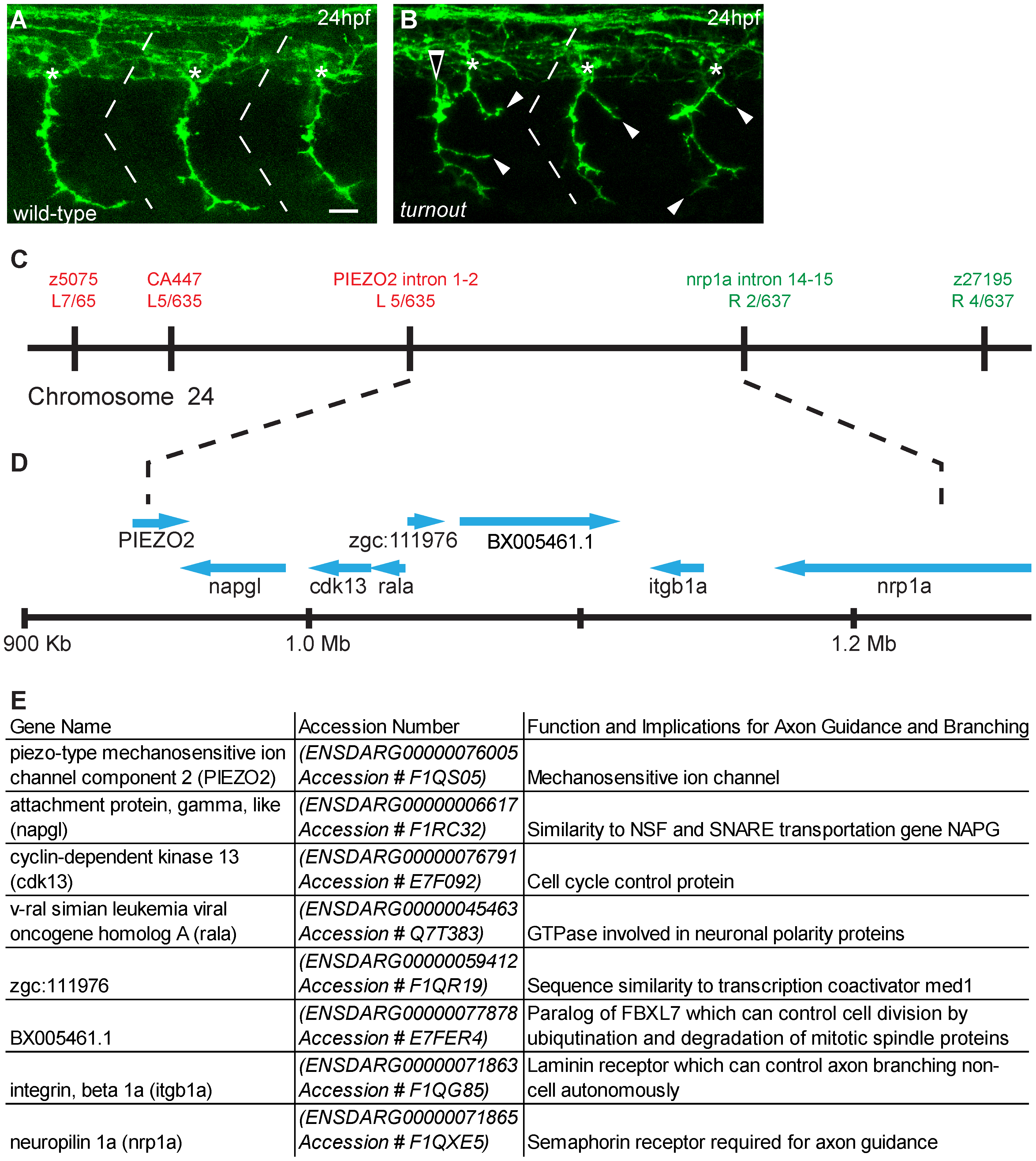
Fig. 1 turnout mutants display guidance and branching defects and maps to chromosome 24 (A) Antibody staining of 24 hpf wild-type embryos to visualize motor axon projections. Note that axons exit the spinal cord at the midsegmental exit point (asterisk) with no branches. (B) In turnout mutants, motor axons exit the spinal cord at ectopic locations (open arrow), and also display long peripheral branches (arrowhead). (C) Genetic map surrounding the turnout locus. Genetic markers and the number of associated recombinant meioses to the left (red text) and to the right (green text) of the turnout mutation are indicated. (D) The physical map of the critical region and annotated genes in this interval. (E) List of annotated genes. Scale bar, 15 µm.