
|
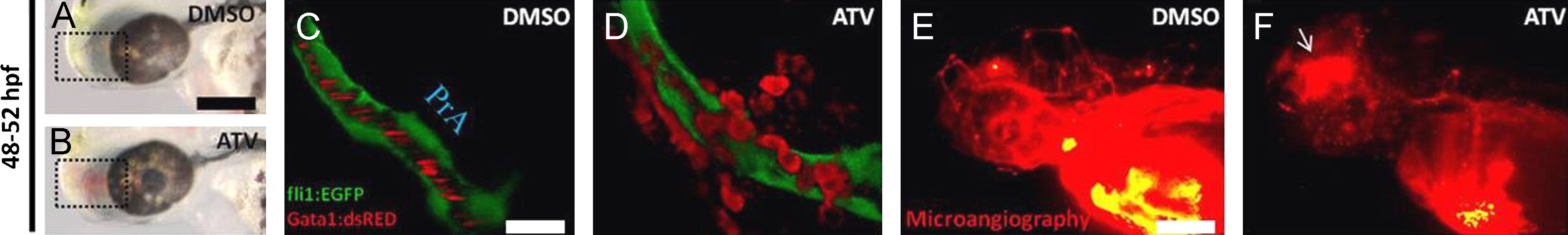
Fig. 3 Pharmacological inhibition of the HMGCR pathway results in loss of vascular stability and induces vessel rupture. ((A) and (B)) Representative bright-field micrographs of 48–52 hpf Tg(fli1:EGFP);(gata-1:DsRed) embryos treated with DMSO or 0.5 mg/L ATV. The boxed region shows where hemorrhage is observed. Scale bar=200 μm. ((C) and (D)) Representative composite confocal Z-stack projections of the boxed regions in the same Tg(fli1:EGFP);(gata-1:DsRed) embryos. Abbreviations: PrA, prosencephalic artery. Scale bar=40 μm. ((E) and (F)) Representative photomicrographs of 48–52 hpf embryos treated with DMSO or 0.5 mg/L ATV and subjected to fluorescence microangiography by intravascular injection of red-fluorescent microspheres. The white arrow shows the site of microsphere extravasation.
Reprinted from Developmental Biology, 373(2), Eisa-Beygi, S., Hatch, G., Noble, S., Ekker, M., and Moon, T.W., The 3-hydroxy-3-methylglutaryl-CoA reductase (HMGCR) pathway regulates developmental cerebral-vascular stability via prenylation-dependent signalling pathway, 258-266, Copyright (2013) with permission from Elsevier. Full text @ Dev. Biol.