
Fig. 1
- ID
- ZDB-IMAGE-130318-33
- Genes
- Publication
- Qian et al., 2013 - ENC1-like Integrates the Retinoic Acid/FGF Signaling Pathways to Modulate Ciliogenesis of Kupffer's Vesicle during Zebrafish Embryonic Development
- All Figures
- Figures for Qian et al., 2013
|
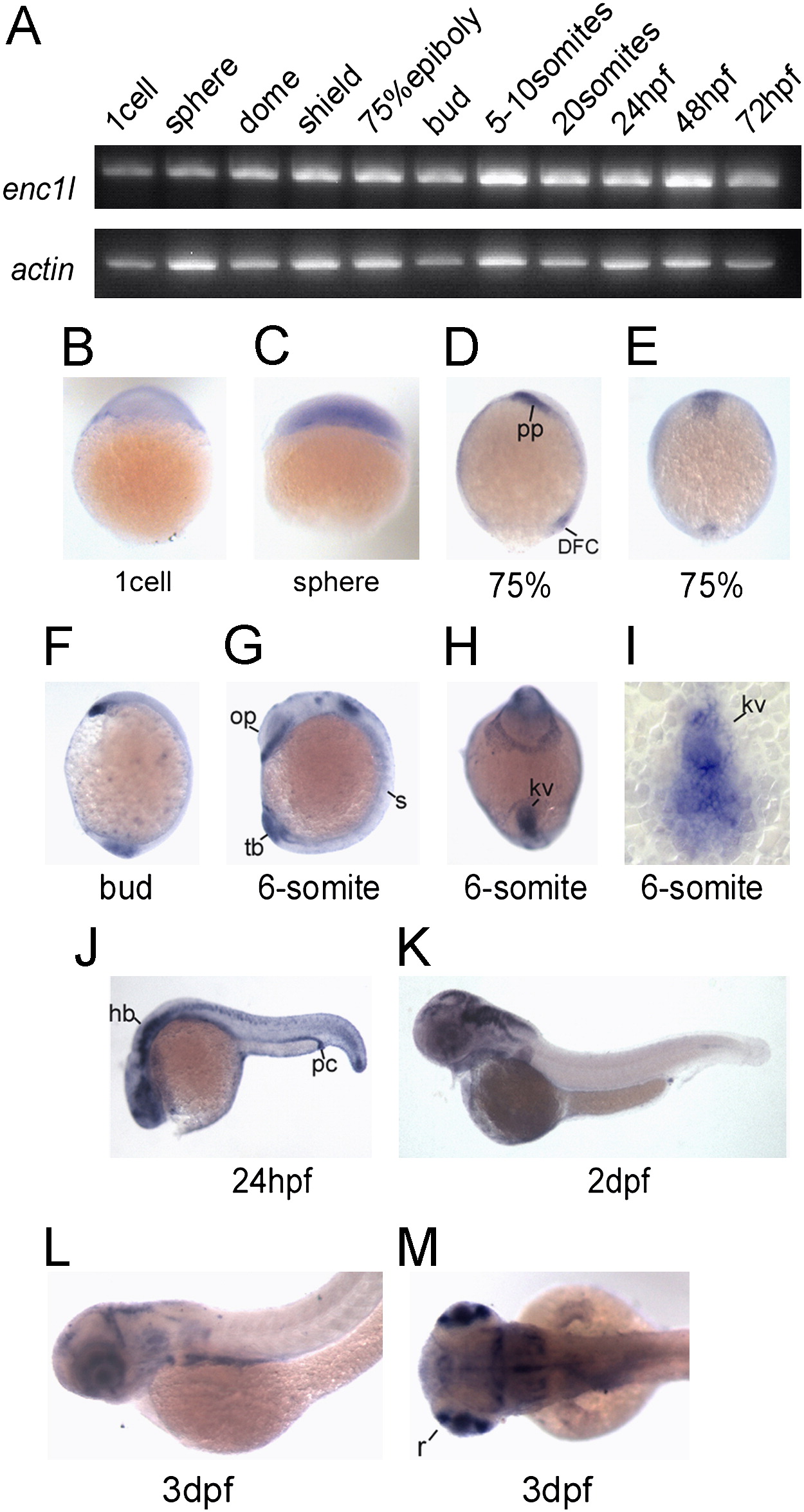
Fig. 1 Enc1l expression pattern analysis in zebrafish. (A) RT-PCR reveals that enc1l is expressed throughout embryonic developmental stages. (B–M) In situ hybridization shows the expression pattern of enc1l during embryonic development. Images of (B) the 1-cell stage and (C) sphere stage show that enc1l is ubiquitously distributed before the gastrula stage. (D, E): In 75% epiboly, enc1l is expressed in the prechordal plate and dorsal forerunner cells. (F): enc1l is expressed in the prechordal plate and the tail bud in the bud stage. (G–I): enc1l is expressed in the 6-somites stage. (G): enc1l is expressed in developing somites, otic primordium, and the tail bud. (H): A ventral view shows that enc1l is expressed in the Kupffer’s vesicle (KV). (I): A dorsal view of a flat-mounted tailbud shows that enc1l is expressed in the KV. (J): In 24hpf, enc1l is mainly expressed in the hindbrain, spinal cord, optic vesicles, and pronephric duct. (K): In 2dpf, enc1l is mainly expressed in the brain and retina. (L, M): enc1l is expressed in retina, brain, and endodermal organs at 3dpf. pp: prechordal plate; kv: Kupffer’s vesicle; DFC: dorsal forerunner cells; s: somites; tb: tail bud; op: otic primordium; pc: pronephric duct; hb: hind brain; r: retina; sc: spinal cord.
Reprinted from Developmental Biology, 374(1), Qian, M., Yao, S., Jing, L., He, J., Xiao, C., Zhang, T., Meng, W., Zhu, H., Xu, H., and Mo, X., ENC1-like Integrates the Retinoic Acid/FGF Signaling Pathways to Modulate Ciliogenesis of Kupffer's Vesicle during Zebrafish Embryonic Development, 85-95, Copyright (2013) with permission from Elsevier. Full text @ Dev. Biol.