
Fig. 7
- ID
- ZDB-IMAGE-130304-8
- Publication
- Meyers et al., 2012 - beta-catenin/Wnt signaling controls progenitor fate in the developing and regenerating zebrafish retina
- All Figures
- Figures for Meyers et al., 2012
|
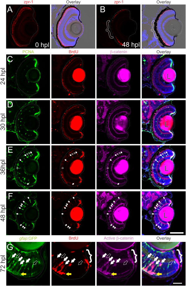
Fig. 7 Wnt signaling is activated as Müller glia reenter the cell cycle following intense-light-induced destruction of photoreceptors.Tg(gfap:GFP)mi2002 zebrafish larvae were exposed to intense light at 6 dpf to lesion their photoreceptors. Immediately following exposure, the retina appeared normal, with photoreceptors (double cones labeled with zpr-1) throughout the ONL (A). Within 48 hours post lesion (hpl), photoreceptors are missing from central retina as shown by the lack of zpr-1 staining (B; bracket). (C-F) Light-lesioned fish were incubated continuously in 2.5 mM BrdU and fixed at 24, 30, 36, or 48 hpl. Cells in the INL become PCNA-positive between 24 and 30 hpl, and begin to incorporate BrdU around 36 hpl (C-E). Immunoreactivity for β-catenin is absent from the INL at 24 or 30 hpl (C, D), but begins to accumulate by 36 hpl (E), when all BrdU-positive, PCNA-positive cells (white arrows) and some BrdU-negative, PCNA-positive cells (open arrows) are immunoreactive. By 48 hpl, all of the PCNA-positive cells are also BrdU-positive and β-catenin-positive (F; white arrows). At 72 hpl, BrdU-positive cells in the INL also express GFP, indicating their Müller glial origins, and they are co-labeled with an antibody for dephosphorylated (active) β-catenin (white arrows; G). Proliferative cells are strongly immunoreactive for dephosphorylated β-catenin (open arrow). The cells of the CMZ also exhibit strong immunoreactivity for dephosphorylated β-catenin (bracket). Scale bars A-F: 100 µm; G: 50 µm.