
|
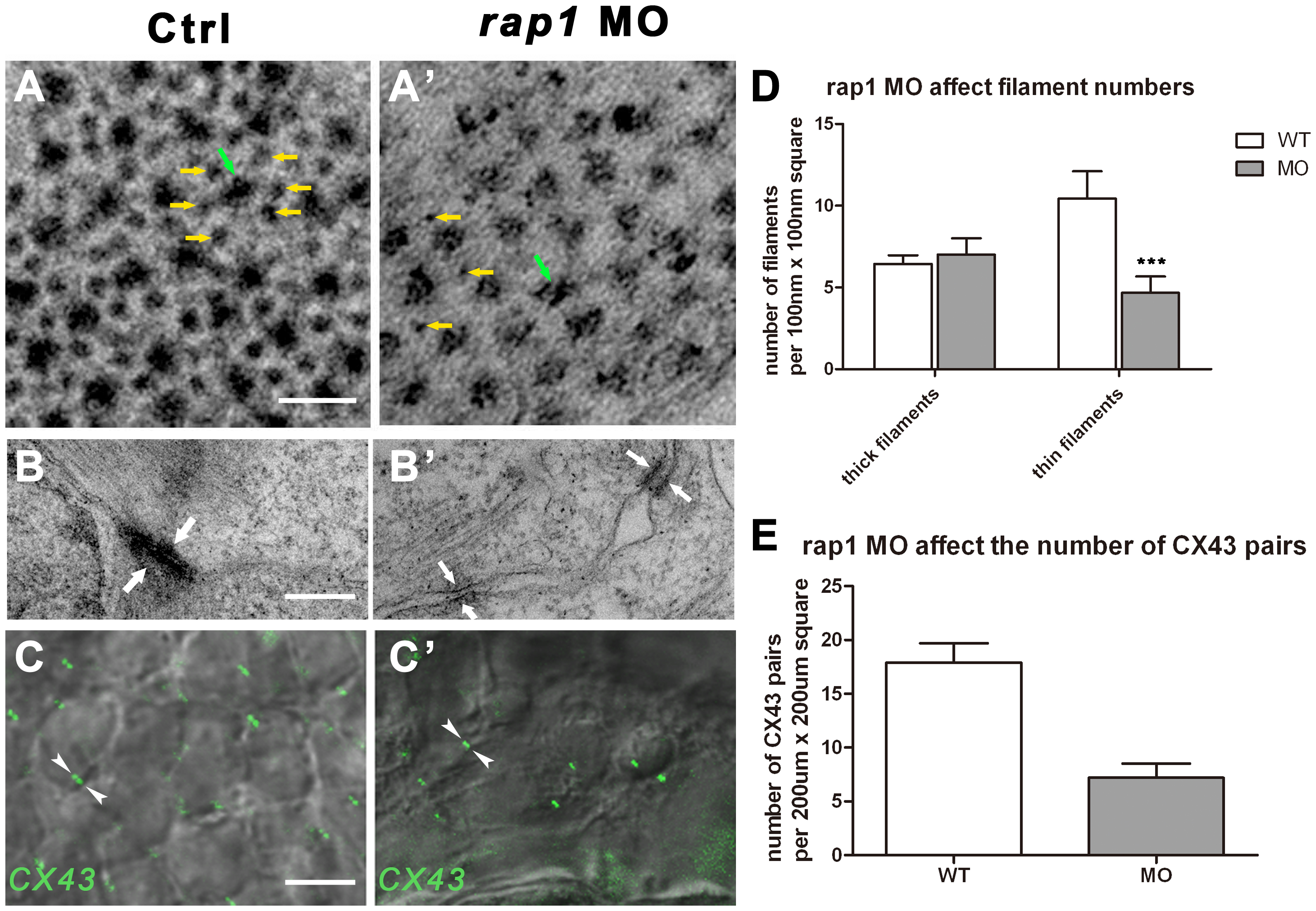
Fig. 3 Ultra-structural changes of cell junctions and sarcomeres in Rap1 knock-down zebrafish heart.
Transmission electronic scan revealed that the myofibril units, regularly organized into hexagonal lattices with thick (green arrow) and thin (yellow arrows) filaments (A) with adherens junctions (AJs) along the membrane of adjacent cardiac myocytes (B). In rap1MO heart, thick filaments (green arrow) number was not markablely changed but thin filaments (yellow arrows) number was significantly and statistically reduced (A′ and D), and less myofibrils attached to AJs (arrows in B and B′), with lager space between the membrane of adjacent cardiac myocytes (B′). Antibody against Connexin 43 stained a significantly and statistically less GJ signal (arrowheads in C and C′) in rap1MO cardiomyocytes at 3 dpf, compared to a normal heart (C, C′, and E). Scale bar, 50 nm in A and A′; 350 nm in B and B′; 50 µm in C and C′. Columns and error bars in D and E showed mean±S.D. n = 9 in filaments experiment (D) for each group, and n = 10 in C×43 antibody staining experiment (E) for each group. Unpaired two-tailed t-test was used to test the significance between two columns in each group in D. *** in statistic graph represent p<0.001 in the t-test.