
Fig. 4
- ID
- ZDB-IMAGE-130225-16
- Publication
- Feng et al., 2012 - The Stress-Response Gene redd1 Regulates Dorsoventral Patterning by Antagonizing Wnt/β-catenin Activity in Zebrafish
- All Figures
- Figures for Feng et al., 2012
|
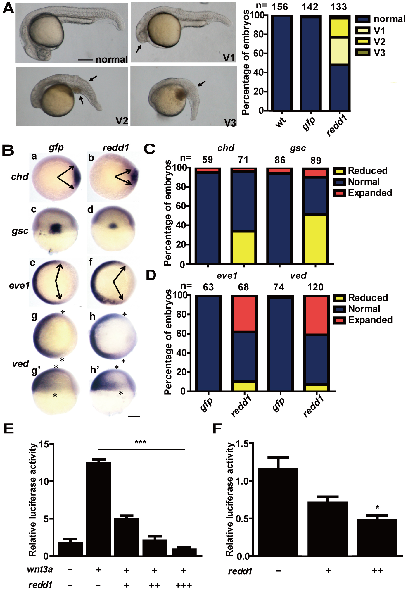
Fig. 4 Redd1 ventralizes embryos and inhibits Wnt signaling.
A) Left panel: classification of phenotypes embryos caused by forced expression of Redd1. 1–2 cell stage embryos were injected with redd1 mRNA. They were raised to 24 hpf and examined. The percentage of embryos in each category is shown in the right panel. B–D) Effects of Redd1 expression. Embryos described in A) were analyzed by whole mount in situ hybridization using the indicated probes. Representative images are shown in B). Panels a–b and e–h are animal pole views with dorsal to the right; panels c–d are dorsal views with animal pole up; panels g2–h2 are lateral views with dorsal to the right and animal pole up. Arrows indicate the edges of the chd and eve1 mRNA expression domains. Asterisks indicate the edges of the ved mRNA expression domain. Scale bar = 200 µm. The percentage of embryos in each category is calculated and shown in C) (chd and gsc) and D) (eve1 and ved). The results are from three independent experiments and the total embryo number is given at the top. E) Redd1 inhibits Wnt3a activity. One-cell stage embryos were injected with 20 pg wnt3a mRNA alone or co-injected with 10 pg (+), 50 pg (++), or 100 pg (+++) redd1 mRNA. TCF/LEF-luciferase reporter DNA was injected in all groups. The embryos were raised to the shield stage and the luciferase activity was determined. Values are means ± S.E. (n = 3). *** P<0.001. F) Redd1 inhibits endogenous Wnt signaling. Embryos were injected with TCF/LEF-luciferase reporter DNA without and with 10 pg (+) or 100 pg (++) redd1 mRNA. The embryos were raised to the shield stage and the luciferase activity was measured. Values are means ± S.E. (n = 3). * P<0.05 compared to the TCF/LEF control group.