
|
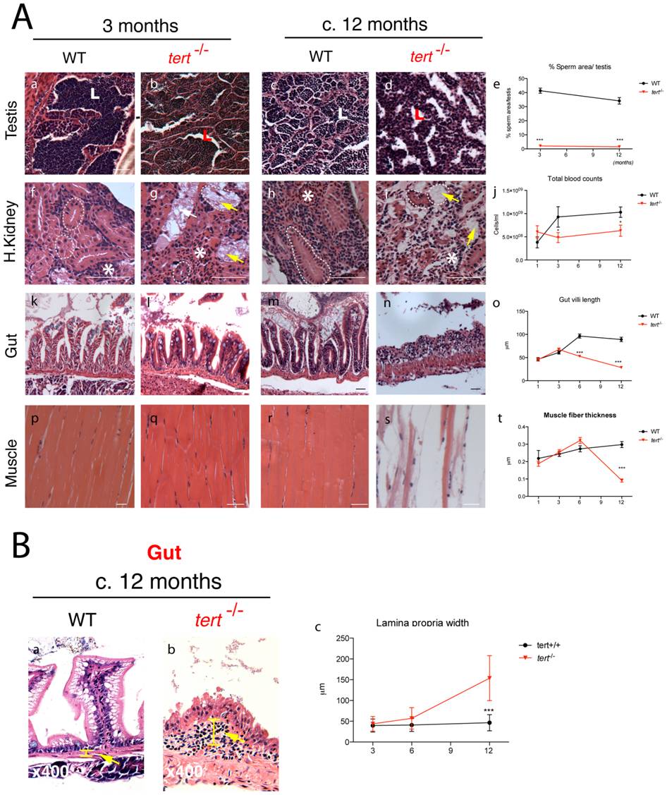
Fig. 3 Telomerase depletion leads to a time- and tissue-dependent degeneration.
A) Representative images of tissue sections of tert-/- Zebrafish and tert+/+ siblings, stained with hematoxilin-eosin. tert-/- zebrafish show progressive tissue deterioration. Severe histological abnormalities are first evident in proliferative tissues (testes, gut and head kidney marrow) and later in non-proliferative (muscle). tert-/- Zebrafish show reduced sperm in testes lumen (L) (Ab and d; p<0.001). The head kidney shows progressive defects in the marrow area (white asterisks) (Ag and i, which correlates with a decrease in total blood (p = 0.0228) cells when compared to tert+/+ siblings from an early age (3 months, Ne5) (Aj). Mesonephric tubules in the head kidney also degenerate in tert-/- (Ag and i dashed outlines). Gut atrophy in tert-/-, reflected as decreased villi length (Al, n, o), becomes significant from the age of 6 months (p<0.001). Muscle fibres are significantly thinner (p<0.001) at terminal time-points (c.12 months) (Aq, s, t, dashed outline); N≥5. B) tert-/- display progressive thickening of gut lamina propria, indicative of inflammation (Bb, c, yellow bar and arrow, Ne4). Data are represented as mean +/- SEM. Scale bar = 50 μm.