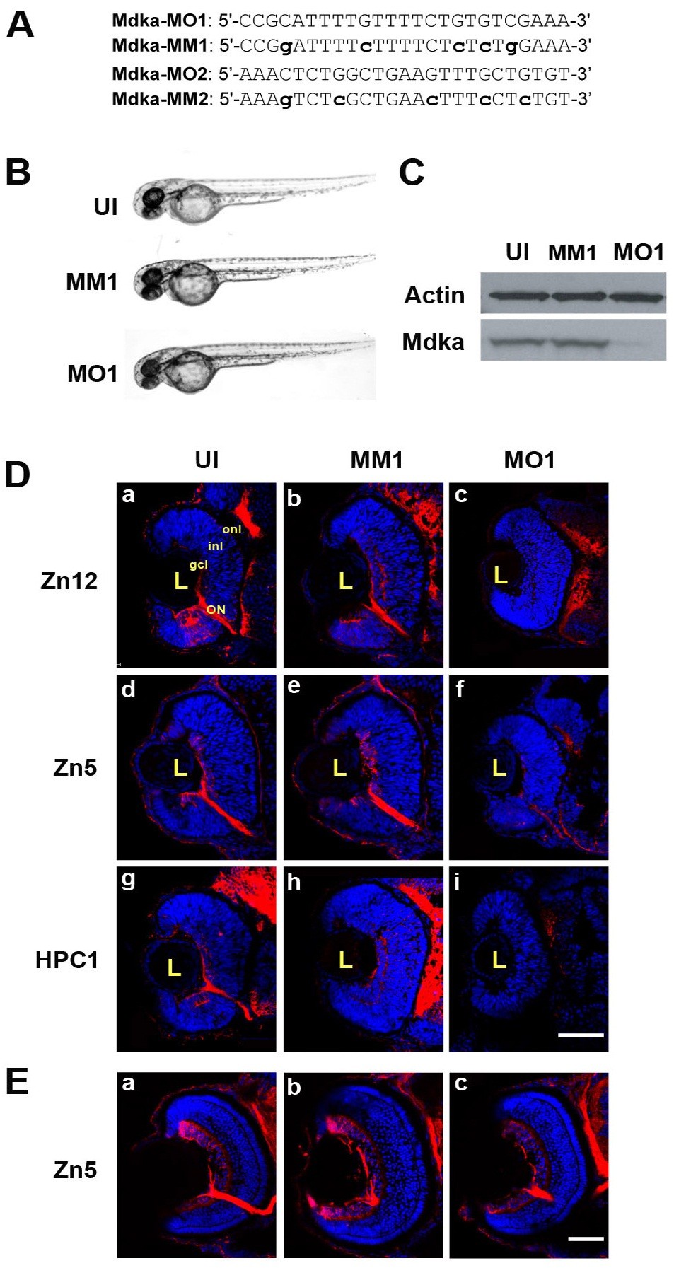
Fig. 1 Mdka -targeted morpholinos inhibit Mdka synthesis and delay neuronal differentiation. (A) Sequences of the two morpholino oligos (MO1, MO2) and the respective mismatch (MM) controls used in this study. (B) Representative embryos from the control and experimental groups at 48 hours post fertilization (hpf). (C) Western blot prepared from 48 hpf embryos. MM, embryos injected with 5-nucleotide mismatch control morpholinos; MO, embryos injected with MO1 Mdka- targeted morpholinos; UI, uninjected control. Note the absence of protein in embryos injected with the mdka-targeted morpholinos (similar results were observed using MO2.) The upper bands are stained with antibodies against actin and serve as loading controls. (D) (a, d, g) Sections taken through the retinas of uninjected embryos (UI) at 48 hpf and stained with antibody markers for ganglion cells (zn12; zn5) and amacrine cells (HPC-1), respectively. (b, e, h) Sections of retinas of embryos injected with 5-nucleotide mismatch morpholinos (MM1) and immunostained as above. (c, f, i) Retinas of embryos injected with Mdka translation-blocking morpholinos (MO1) and immunostained as above. Note the absence of differentiation markers that label inner retinal neurons. (E) Representative sections from uninjected (a), control (b), and loss-of-function embryos (c) allowed to survive to 72 hpf. The absence of neuronal differentiation at 48 hpf is recovered by 72 hpf. gcl, ganglion cell layer; inl, inner nuclear layer; L, lens; ON, optic nerve; onl, outer nuclear layer; . Scale bar equals 50μm. MIP 3D; Maximum intensity projection, three dimensional.
Image
Figure Caption
Figure Data
Acknowledgments
This image is the copyrighted work of the attributed author or publisher, and
ZFIN has permission only to display this image to its users.
Additional permissions should be obtained from the applicable author or publisher of the image.
Full text @ Neural Dev.

