
Fig. 10
- ID
- ZDB-IMAGE-130218-33
- Publication
- Janssens et al., 2013 - Matrix metalloproteinase 14 in the zebrafish: an eye on retinal and retinotectal development
- All Figures
- Figures for Janssens et al., 2013
|
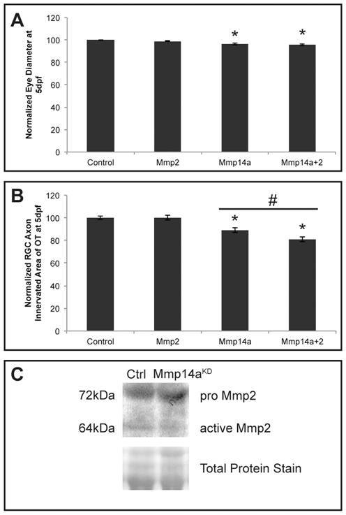
Fig. 10 In vivo interaction between Mmp14a and Mmp2.
A, B Quantitative analysis of eye size (A) and tectal area innervated by RGC axons (B) after single or combined suboptimal knockdown of Mmp14a and Mmp2 in 5 dpf embryos reveals that suboptimal knockdown of Mmp2 does not affect eye size nor OT innervation. However, combined knockdown of Mmp14a and Mmp2 results in embryos with normal eye size but a significantly reduced RGC axon innervation area in the OT, as compared to the Mmp14a suboptimal knockdown embryos (n = 75 from 3 independent experiments). C Western blot for Mmp2 on extracts of control and Mmp14a morphant embryos at 30 hpf shows reduced levels of active Mmp2 protein (64 kDa) in Mmp14a morphant embryos. Total protein coomassie blue staining was used for loading control. Data are represented as mean ± SEM (*p<0.05 versus controls using a Student′s t-test; #p<0.05 between 1 ng injections of Mmp14a-MO and Mmp14a+Mmp2-MO using a multilevel model statistical test (SAS proc mixed)). dpf: days post fertilization; hpf: hours post fertilization; MO: morpholino; OT: optic tectum; RGC: retinal ganglion cells.