
Fig. 4
- ID
- ZDB-IMAGE-130218-17
- Publication
- Nikaido et al., 2013 - A Systematic Survey of Expression and Function of Zebrafish frizzled Genes
- All Figures
- Figures for Nikaido et al., 2013
|
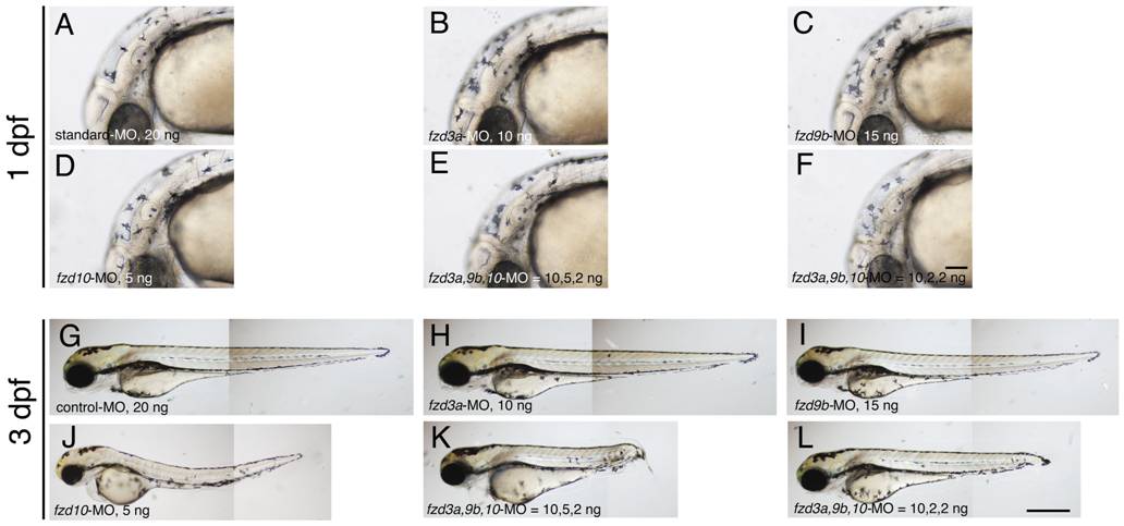
Fig. 4 Morpholino-mediated knockdown of fzd3a, fzd9b and fzd10 in the sensitized background.
A–F) 1 dpf embryos. Melanisation patterns of typical embryos showing “elongated axis + no/slight necrosis” are presented here (see Table 4 for quantitation). G–L) 3 dpf embryos. All are left side views with dorsal oriented to the top. A, G) standard Control-MO, 20 ng/embryo. B, H) fzd3a-MO injected. 10 ng/embryo. C, I) fzd9b-MO injected. 15 ng/embryo. D, J) fzd10-MO injected. 5 ng/embryo. E, K) fzd3a, fzd9b, fzd10-MO = 10, 5, 2, ng/embryo, respectively. F, L) fzd3a, fzd9b, fzd10-MO = 10, 2, 2, ng/embryo, respectively. Scale bars: (A–F) 100 μm, (G–L) 500 μm.