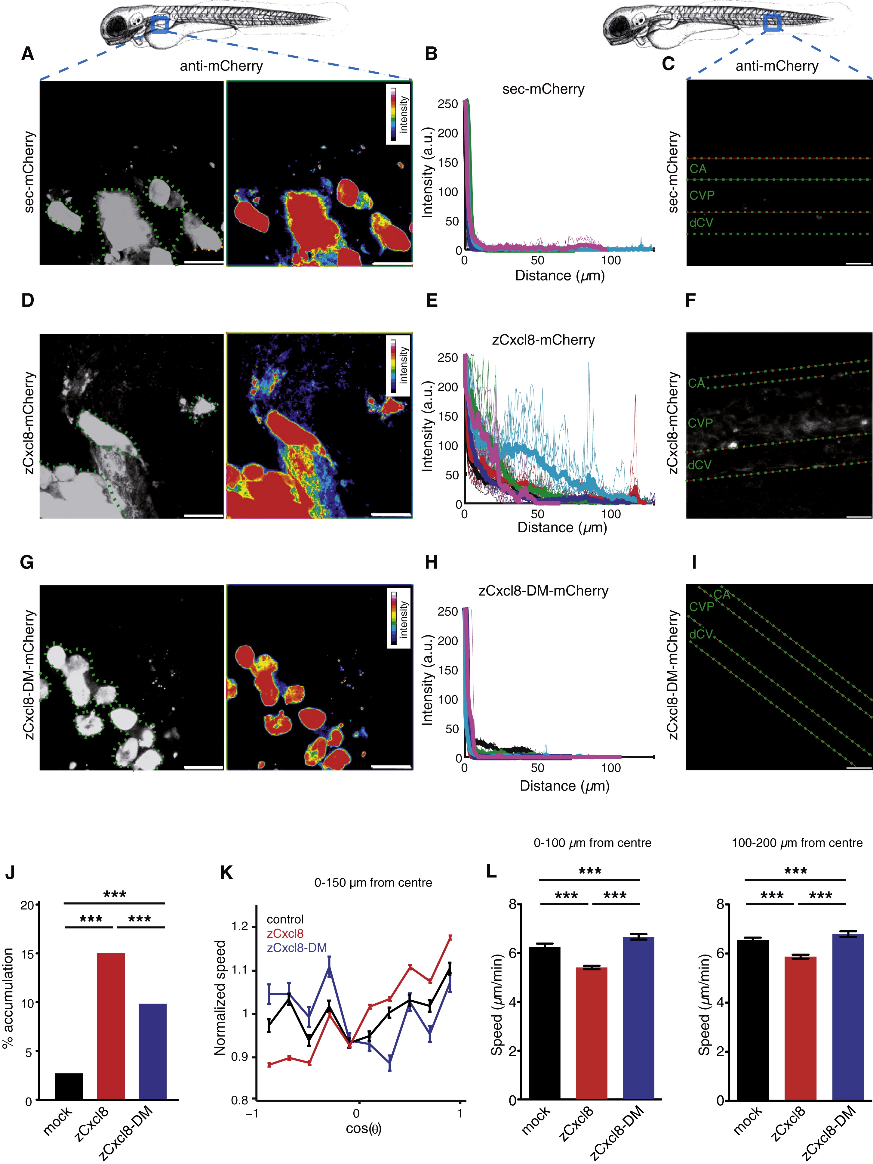
Fig. 4 zCxcl8 Directs and Restricts Neutrophil Motility as HSPG-Bound Gradients(A–I) Extracellular distribution of sec-mCherry (A–C), zCxcl8-mCherry (D–F), and zCxcl8-DM-mCherry (G–I) secreted from transplanted cells. Top: 3 dpf larvae. Squares indicate imaged regions.(A, D, and G) Projections of confocal image stacks showing immunodetection of sec-mCherry, zCxcl8-mCherry, and zCxcl8-DM-mCherry at the site of secretion. Left: grayscale images with green dotted lines outlining the secreting cells. Right: same images color coded for fluorescence intensity.(B, E, and H) Quantification of mCherry staining as a function of the distance from the nearest producing cell. Fluorescence intensity signals along various lines starting from the implants is shown. Colors are associated to different larvae, thin curves are the intensity evolutions along individual lines, and thick curves are the average of the thin curves. Data are from six or seven larvae in two or three independent experiments.(C, F, and I) Projections of confocal image stacks showing immunodetection of sec-mCherry, zCxcl8-mCherry, and zCxcl8-DM-mCherry in the vasculature. CA, caudal artery; CVP, caudal vein plexus; dCV, definitive caudal vein. Data are representative of six or seven larvae in two or three independent experiments. For (A), (C), (D), (F), (G), and (I), scale bars represent 25 μm.(J) Percentage of neutrophil tracks reaching transplanted cells expressing or not expressing zCxcl8 or zCxcl8-DM. Mock: n = 568; zCxcl8: n = 1,378; zCxcl8-DM: n = 772 (Fisher′s exact test for tracks reaching or not transplant). p < 0.0001.(K) Evolution of the average normalized speed with the orientation of motion (cosine of the angle θ, with θ = 0 the direction of the transplant center) within a radius of 150 μm from the transplant center. Speed values are normalized by the average value of the speed, to allow comparison across different experiments. Data are from four larvae in two to four independent experiments. Error bars indicate SEM.(L) Average speed of neutrophil steps inside a radius of 100 μm (left) and between radii of 100 μm and 200 μm (right). Mock: n = 2,293 (left) and n = 6,924 (right); zCxcl8: n = 9,208 (left) and n = 14,375 (right); zCxcl8-DM: n = 3,269 (left) and n = 6,589 (right). Data are from four larvae in two to four independent experiments (Mann-Whitney test). Error bars indicate SEM. p < 0.0001.See also Figure S4 and Movie S4.
Image
Figure Caption
Acknowledgments
This image is the copyrighted work of the attributed author or publisher, and
ZFIN has permission only to display this image to its users.
Additional permissions should be obtained from the applicable author or publisher of the image.
Full text @ Curr. Biol.

