
IMAGE
Fig. S1
- ID
- ZDB-IMAGE-130205-10
- Publication
- Huitema et al., 2012 - Entpd5 is essential for skeletal mineralization and regulates phosphate homeostasis in zebrafish
- All Figures
- Figures for Huitema et al., 2012
Image
|
Figure Caption
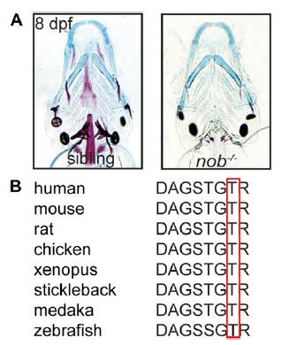
Fig. S1 (A) Alcian blue staining is unaltered in nob mutants versus siblings. The images are similar to the ones presented in Fig. 1A, but taken with higher contrast settings to allow an appreciation of normal chondrocyte morphology in mutant embryos. (B) Multiple sequence alignment of Entpd5 proteins demonstrating the conserved nature of zebrafish Thr80 in the first apyrase domain.
Acknowledgments
This image is the copyrighted work of the attributed author or publisher, and
ZFIN has permission only to display this image to its users.
Additional permissions should be obtained from the applicable author or publisher of the image.
Full text @ Proc. Natl. Acad. Sci. USA