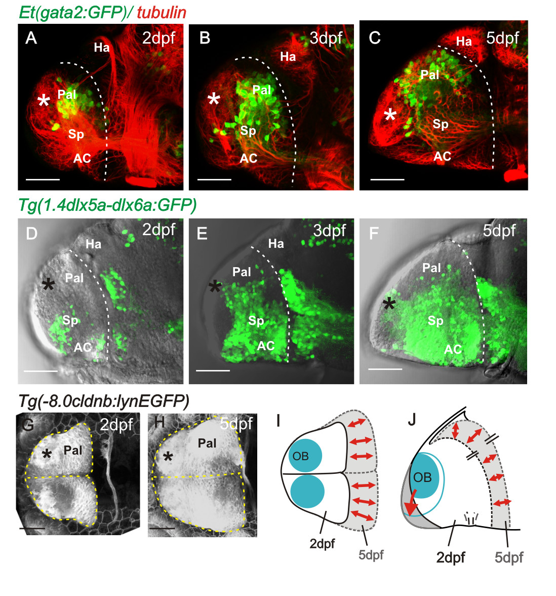
Fig. 4 Pallial and subpallial expansion between 2 dpf and 5 dpf. A to C. Lateral views of 2, 3, and 5 day telencephali expressing [Et(gata2:GFP)bi105] in pallial neurons whose numbers expand into the region between the OB (asterisk) and the diencephalic habenulae. D to F. Lateral views of 2, 3, and 5 day telencephali expressing Tg(1.4dlx5a-dlx6a:GFP)ot1 in subpallial cells, whose numbers expand greatly between days 2 and 5. G and H. Dorsal views of telencephalon at 2 and 5 days labelled with the transgene 8.0cldnb:lynEGFP. Positions of olfactory bulbs are superimposed and the outlines of the telencephali are overlaid in I (dorsal view) and J (lateral view), to illustrate that most growth occurs along the anteroposterior axis but that the mediolateral dimensions alter relatively little. AC: anterior commissure; Hab: habenula; OB: olfactory bulb; Pal: pallium; Sp: subpallium.
Image
Figure Caption
Acknowledgments
This image is the copyrighted work of the attributed author or publisher, and
ZFIN has permission only to display this image to its users.
Additional permissions should be obtained from the applicable author or publisher of the image.
Full text @ Neural Dev.

