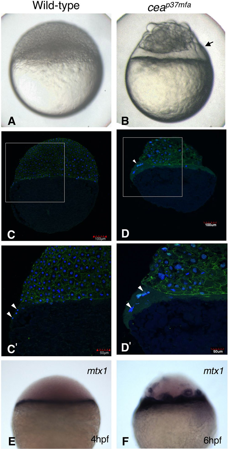
Fig. S1 The cea mutant has an enlarged YSL. (A) Wild-type and (B) cea mutant embryos at 4 hpf. Arrow in B indicates the enlarged syncytial layer. (C–D′) Cryosections of wild-type and mutant embryos at 4 hpf, stained with anti-β-catenin antibody (green) and DAPI (blue). C′ and D′ show enlargements of the areas indicated by squares in C and D, respectively. Nuclei (white arrowheads) are not separated by cell membranes in both wild-type and mutant embryos. The clusters of nuclei and surrounding cytoplasm are larger in the cea mutant than the wild-type embryo (compare C′ with D′). (E,F) Whole-mount in situ hybridization of (E) control and (F) cea mutant embryos with the YSL marker, mtx1. The enlarged syncytial layer expresses mtx1 at 6 hpf.
Image
Figure Caption
Figure Data
Acknowledgments
This image is the copyrighted work of the attributed author or publisher, and
ZFIN has permission only to display this image to its users.
Additional permissions should be obtained from the applicable author or publisher of the image.
Full text @ Biol. Open

