
Fig. 2
- ID
- ZDB-IMAGE-130117-32
- Publication
- Sousa et al., 2012 - A new zebrafish bone crush injury model
- All Figures
- Figures for Sousa et al., 2012
|
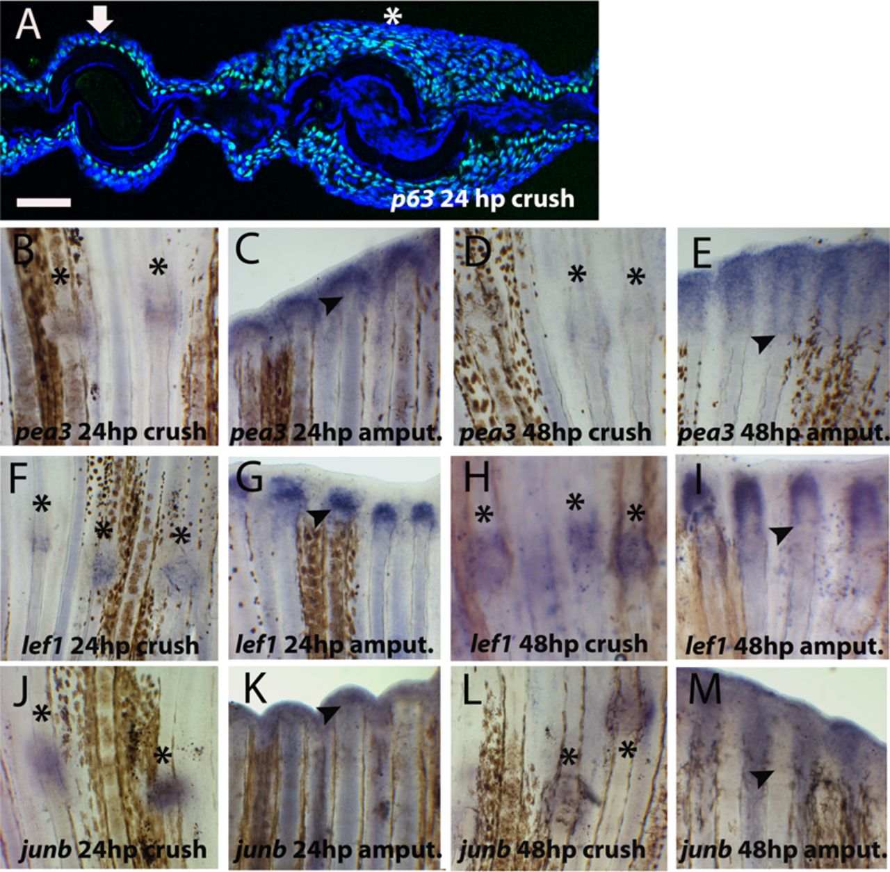
Fig. 2 Presence of wound healing marker genes after crush injury.
(A) Immunohistochemistry for p63 (green) in 24hpc transversal section of caudal fin. Blue represents DAPI positive nuclei. The arrow indicates an intact bony ray and the asterisk the site of crush injury. (B–M) Whole-mount in situ hybridization for mRNA of (B–E) pea3 at (B) 24hpc (C) 24hpa (D) 48hpc (E) 48hpa; (F–I) lef1 at (F) 24hpc (G) 24hpa (H) 48hpc (I) 48hpa; (J–M) junb at (J) 24hpc (K) 24hpa (L) 48hpc (M) 48hpa. Arrowheads indicate the amputation plane and asterisks indicate crush injury sites. Scale bar corresponds to 50μm in A and 200μm in B–M. (hpa – hours post-amputation; hpc – hours post-crush injury).