
|
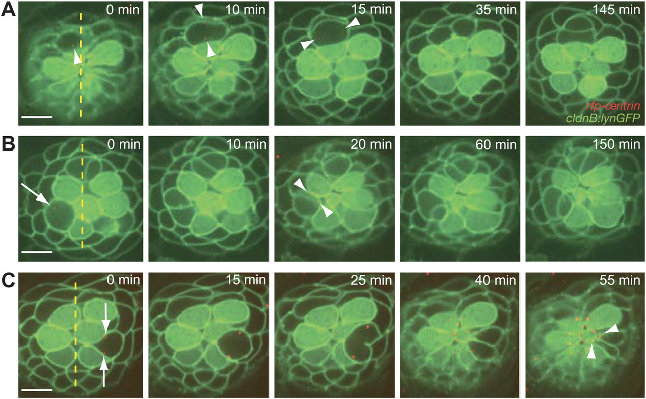
Fig. 2 Normal cellular positioning and orientation in a neuromast.
(A) The centrosomes of a dividing hair-cell precursor, which are marked red with centrin-RFP (arrowheads), rotate 90° to position the axis of division along the neuromast′s axis of polarity. After cytokinesis has been completed, rearrangement between the daughter cells commences. (B) A time-lapse series shows the rearrangement of a pair of hair cells following the division of a precursor orthogonally to a neuromast′s axis of polarity. An arrow in the first panel marks the position of one centrosome in a dividing hair-cell precursor; the other centrosome is out of the plane of focus. Apical movement of the centrosomes (arrowheads) coincides with the rearrangement, after which the hair-cell pair undergoes an additional 90° rotation to align with the neuromast′s axis of polarity. Note the rearrangement of a second pair of nascent hair cells at the dorsal pole of the neuromast. (C) A pair of supporting cells is formed through the symmetric division of a precursor positioned laterally to the central cluster of hair cells. The centrosomes in the precursor (arrows) move to position the axis of division obliquely relative to the neuromast′s axis of planar polarity. Although the centrosomes move apically (arrowheads), the supporting-cell pair does not undergo rearrangements. The scale bars represent 10μm in all panels.