
Image
|
Figure Caption
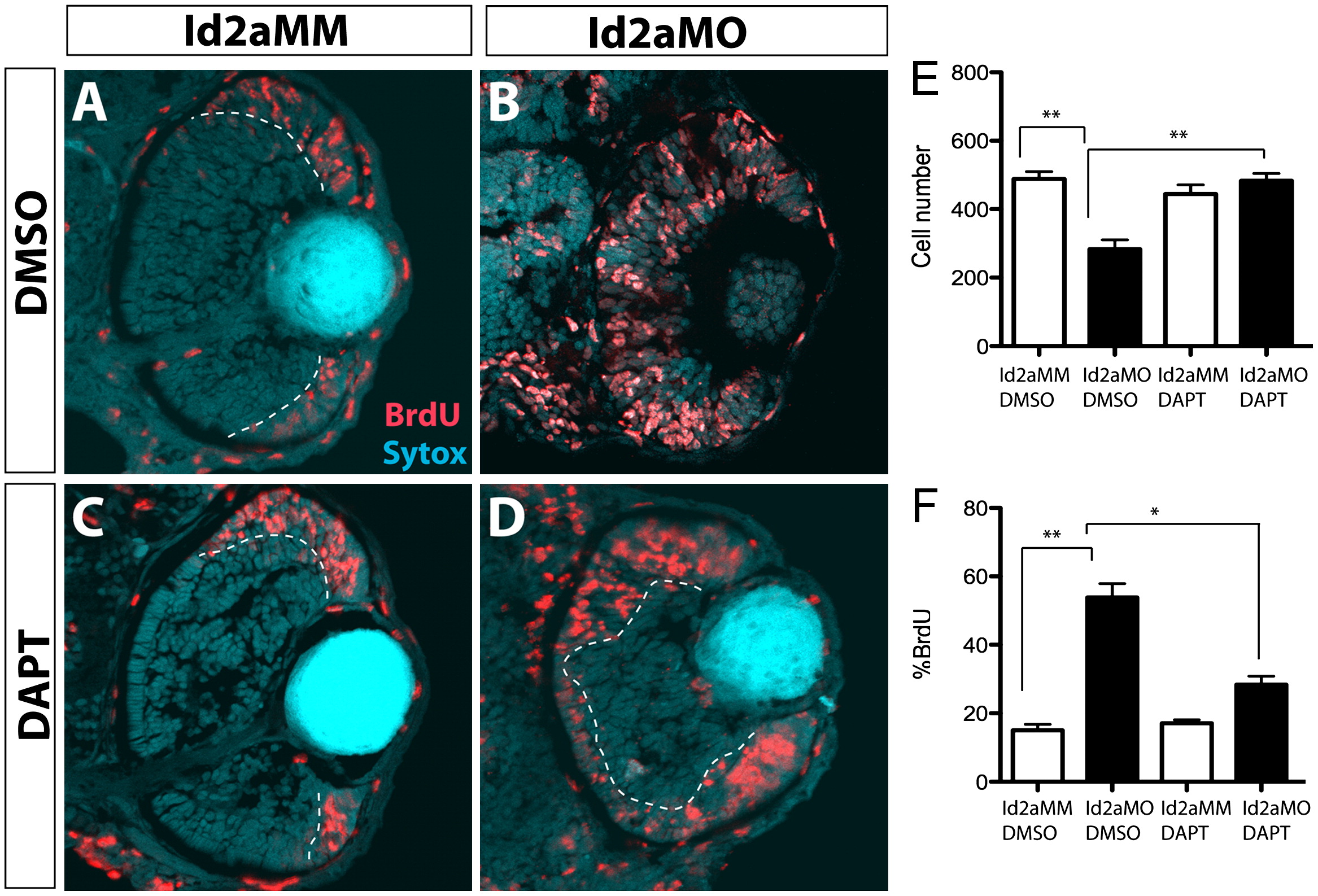
Fig. 1 DAPT inhibition of the Notch pathway rescues proliferation defects in Id2a-deficient retinae. (A)–(D) Id2aMM and Id2aMO embryos treated with either (A) and (B) DMSO or (C) and (D) DAPT from 28–48 hpf and pulsed with BrdU for 30 min prior to fixation. Transverse retinal sections showing BrdU-positive cells at 48 hpf (red). Quantification of average cell number per retinal section (E) and the proportion of BrdU-positive cells per retinal section (F) at 48 hpf. Nuclei are stained with Sytox-green (cyan). Dorsal is up in all images. Error bars represent SEM, n=9; *p<0.05, **p<0.005.
Figure Data
Acknowledgments
This image is the copyrighted work of the attributed author or publisher, and
ZFIN has permission only to display this image to its users.
Additional permissions should be obtained from the applicable author or publisher of the image.
Reprinted from Developmental Biology, 371(2), Uribe, R.A., Kwon, T., Marcotte, E.M., and Gross, J.M., Id2a functions to limit Notch pathway activity and thereby influence the transition from proliferation to differentiation of retinoblasts during zebrafish retinogenesis, 280-292, Copyright (2012) with permission from Elsevier. Full text @ Dev. Biol.