
Fig. 5
- ID
- ZDB-IMAGE-121212-37
- Publication
- Gyda et al., 2012 - The tumor suppressor gene retinoblastoma-1 is required for retinotectal development and visual function in zebrafish
- All Figures
- Figures for Gyda et al., 2012
|
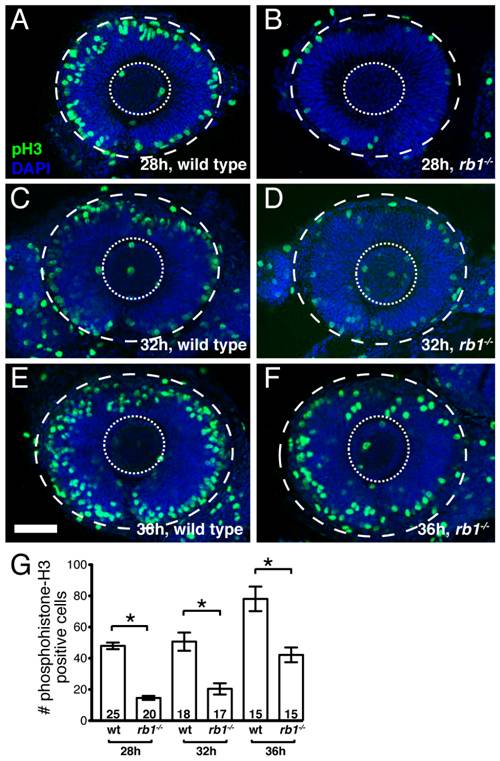
Fig. 5 rb1te226a retinas show delayed cell cycle exit during early retinogenesis.
Retinas removed from wild type (A, C, E) or rb1te226a embryos (B, D, F) at 28 (A-B), 32 (C-D), or 36 hpf (E-F). Retinas labeled with anti-phosphohistone H3 antibody (green) to label M-phase nuclei and counterstained with DAPI (blue). Lateral view of maximum intensity projection of confocal z-stacks. Inner dashed circle outlines lens and outer dashed circle outlines retina. (G) Mean number of anti-phosphohistone-H3 labeled cells per retina. Error bars denote SEM. *p<0.01; one-way ANOVA with brackets indicating compared groups. N retinas shown at base of bar graphs. Anterior to the left, dorsal to the top of each panel. Scale bar = 50 μm.