
Fig. S2
- ID
- ZDB-IMAGE-121212-33
- Publication
- Gyda et al., 2012 - The tumor suppressor gene retinoblastoma-1 is required for retinotectal development and visual function in zebrafish
- All Figures
- Figures for Gyda et al., 2012
|
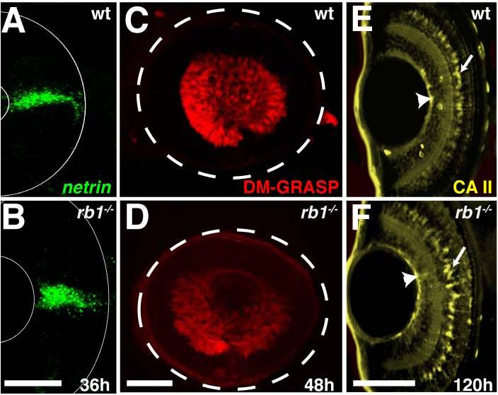
Fig. S2 rb1te226a retinas express markers indicating normal gross morphology. Retinas removed from wild type (A, C, E) or rb1te226a embryos (B, D, F) at 36 (A–B), 48 (C–D), or 120 hpf (E–F). (A,B) netrin in situ shows presence of glial cells at optic stalk of retina. (C, D) DM-GRASP immunolabeling labels postmitotic, differentiated RGCs. White dashed circle outlines retina. (E, F) Carbonic anhydrase II (CA II) immunolabeling identifies Muller glia cell bodies (arrows) and endfeet (arrowheads). Lateral views of maximum intensity projection of confocal z-stacks. Anterior to the left, dorsal to the top of each panel. Scale bar = 50 μm.