
Fig. 1
- ID
- ZDB-IMAGE-121212-12
- Publication
- Pase et al., 2012 - Neutrophil-Delivered Myeloperoxidase Dampens the Hydrogen Peroxide Burst after Tissue Wounding in Zebrafish
- All Figures
- Figures for Pase et al., 2012
|
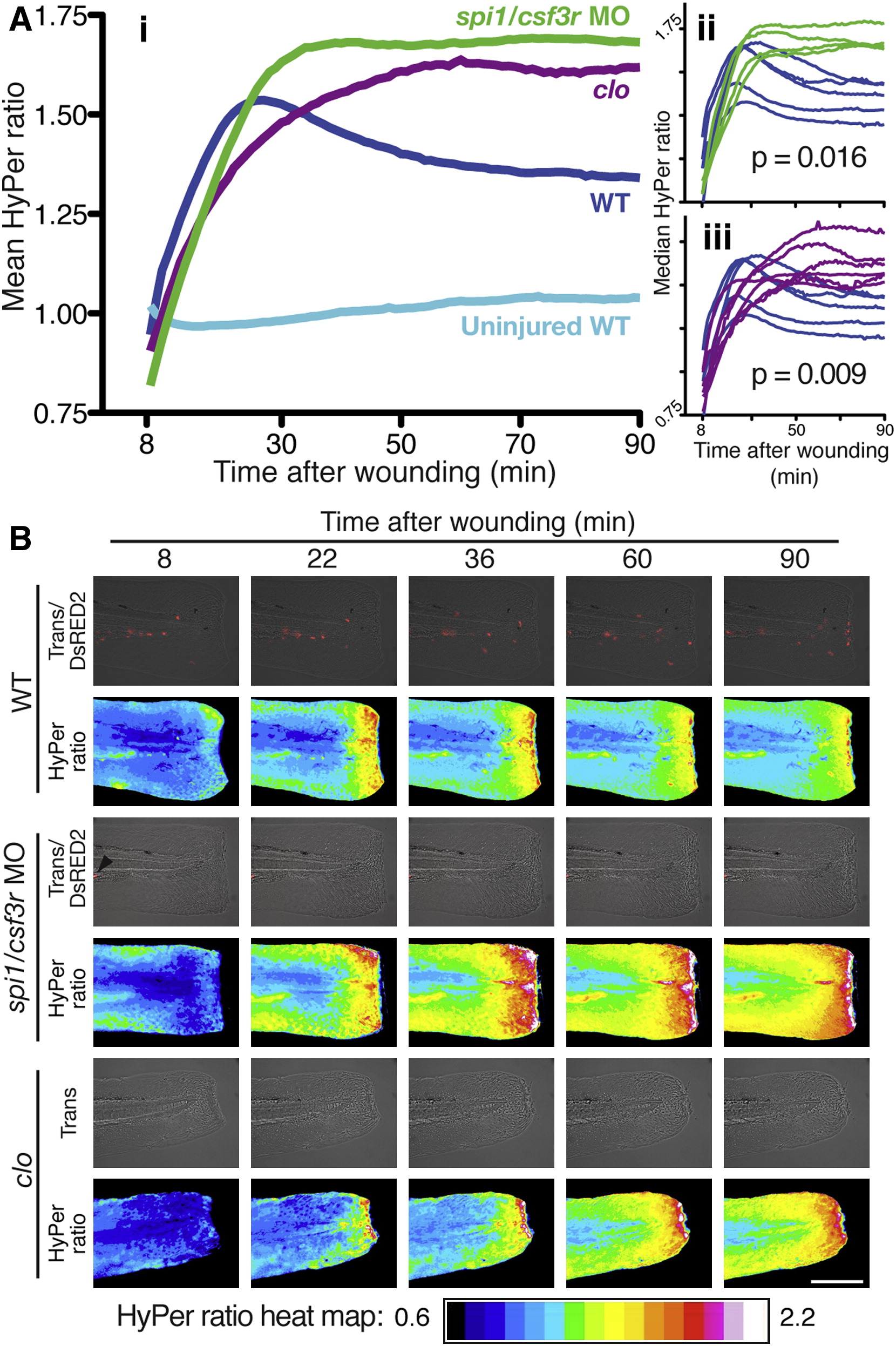
Fig. 1 Leukocytes Are Required for the Initial Decline in Wound Margin H2O2 Concentrations(A) (i) Mean HyPer ratios reflecting the wound zone [H2O2] profile of WT (injured and uninjured), cloche mutant (clo) and spi1/csf3r morphant embryos. Note the significantly sustained, elevated HyPer ratio/[H2O2] in leukocyte-deficient clo and spi1/csf3r morphants. Insets (ii, iii) show the individual median HyPer data and p values (natural permutation test). The n values are as follows (embryos/independent days/scans): injured WT, 5/2/4; spi1/csf3r morphants, 4/2/3; clo 6/3/5; uninjured WT, 5/1/5.(B) HyPer heat maps depicting [H2O2] across the wounded tail fin at five sequential time points. Accompanying overlaid transmission (Trans) and DsRED2 images demonstrate number and position of Tg(lyz:DsRED2) neutrophils. Note the single DsRED2-positive cell in the spi1/csf3r morphant (black arrow head). clo did not carry the lyz:DsRED2 transgene. Scale bar (applies to all panels) represents 200 μm. Heat map legend applies to all figures. The stills are from Movie S1.