
Fig. 5
- ID
- ZDB-IMAGE-121205-7
- Publication
- Blaker-Lee et al., 2012 - Zebrafish homologs of 16p11.2, a genomic region associated with brain disorders, are active during brain development, and include two deletion dosage sensor genes
- All Figures
- Figures for Blaker-Lee et al., 2012
|
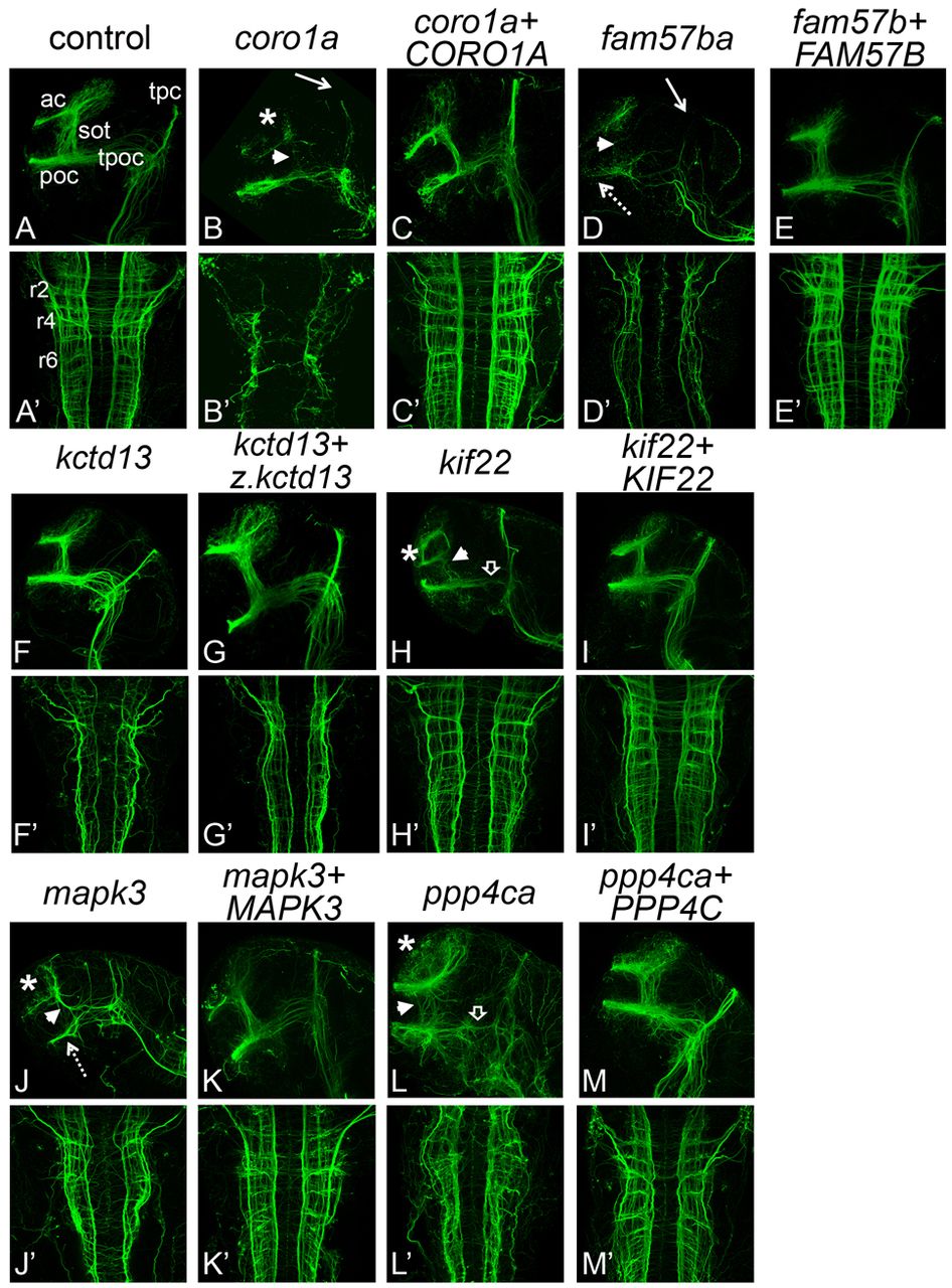
Fig. 5 Axon tracts are abnormal in LOF embryos. Forebrain and hindbrain axon tracts after LOF. Axons were labeled with anti-acetylated α-tubulin antibody and imaged by scanning confocal microscopy of fixed, flat-mounted 36 hpf LOF embryos. (A-M) Lateral view, showing forebrain axons. (A′-M′) Dorsal view, showing hindbrain axons. Genes targeted for LOF by MO injection are indicated above each set of panels in lowercase. Over two independent experiments, an average of eight embryos per gene were imaged for effects of LOF and rescue. The percentage of affected embryos was 80% or greater. Hindbrain and forebrain tracts were affected in all LOF conditions, except kctd13, which only showed defects in the hindbrain, and kif22, which only showed defects in the forebrain. The rescues with cognate RNA led to rescue in 75–100% of embryos assayed. Human RNAs co-injected for rescue are indicated by uppercase letters, except for z.kctd13, which refers to RNA from the zebrafish gene. ‘Control’ embryos were injected with control MO (see Methods). ac, anterior commissures; sot, supra optic tract; poc, post optic commissure; tpoc, tract of the post optic commissure; tpc, tract of the posterior commissure; r2, r4, r6, rhombomeres 2, 4, and 6; asterisk, reduced or disorganized ac; white arrowhead, reduced or disorganized sot; white arrow, reduced tpc; open arrowhead, reduced or disorganized tpoc; dotted arrow, reduced poc.