
|
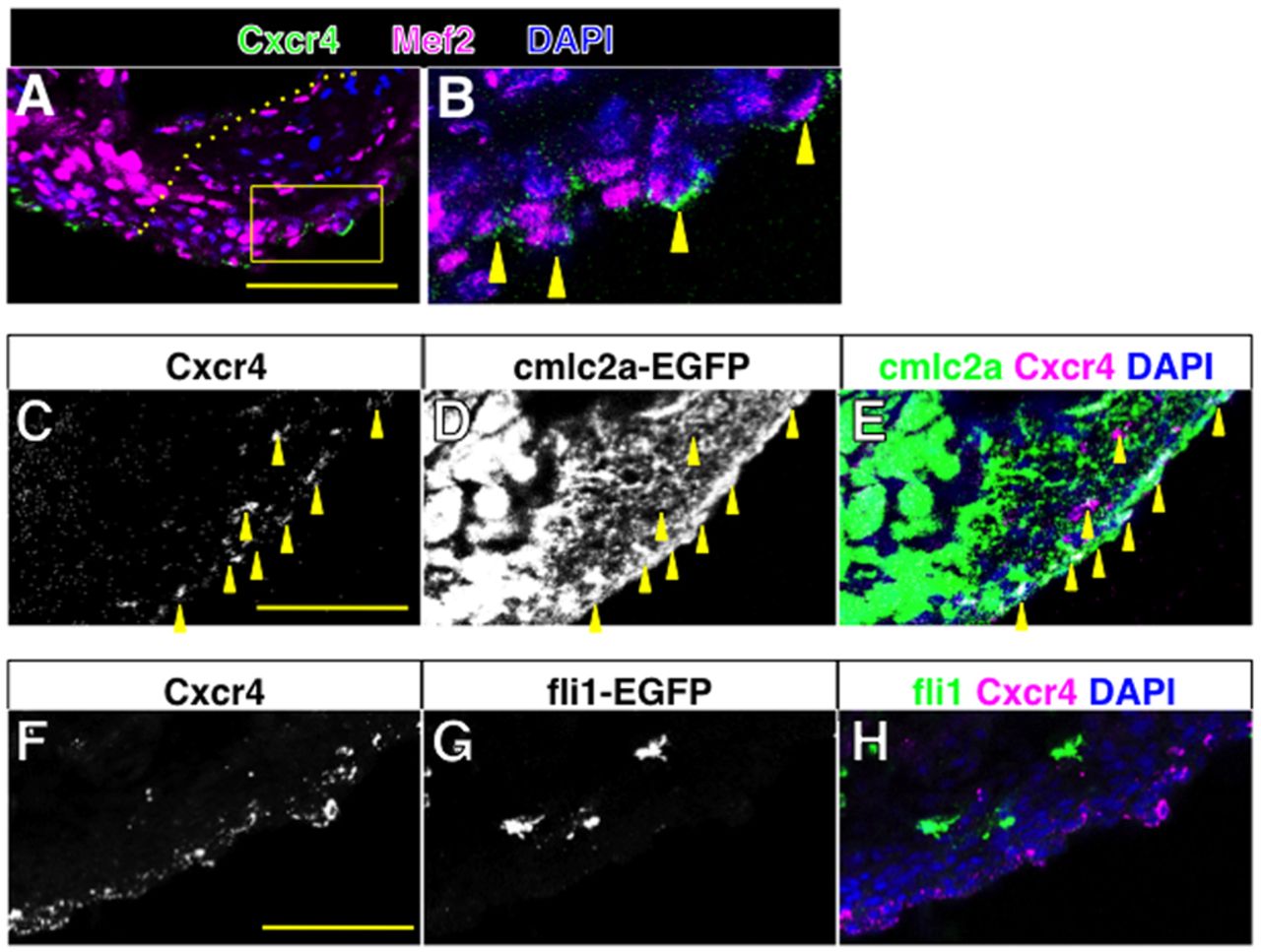
Fig. 2 Cxcr4 is present on CMs. (A,B) Confocal images of Cxcr4 (green) and Mef2 (magenta) double staining of control heart at 7 dpa. The Cxcr4 signal was detected at the cell surface of nuclear Mef2-positive cells in the regenerating area (arrowheads in B). B shows a higher magnification image of the boxed area in A. The dotted line indicates the amputation plane. (C-E) Confocal images of Cxcr4 (C) and cmlc2a-EGFP signal, detected by anti-GFP antibody (D), and a merged image (E). The arrowheads point to cells positive for both Cxcr4 and cmlc2a-EGFP signals. (F-H) Confocal images of Cxcr4 (F) and fli1-EGFP signal, detected by anti-GFP antibody (G), and a merged image (H). Scale bars: 50 μm.