
|
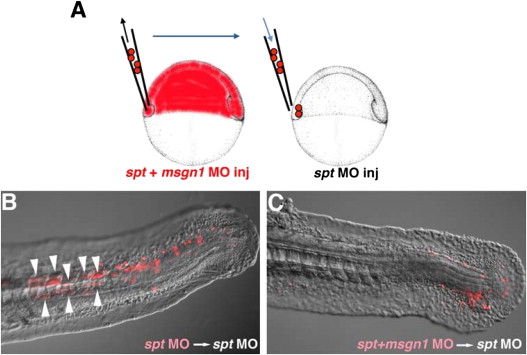
Fig. 5 Msgn1 functions in a cell-autonomous manner in tail PSM formation. (A) Schematic representation of the transplantation experiment. Rhodamine-labeled cells from spt MO- or msgn1;spt MO-injected embryos were transplanted to the ventral margin of the spt MO-injected host embryo at the shield stage. (B and C) While the spt single knocked down cells could contribute to the formation of the tail PSM and differentiation of the tail muscle in the host embryo (B, n=34; 71%), spt and msgn1 double knocked down cells remained at the posterior tip of the tail and failed to contribute to formation of the tail muscle (C, n=32; 0%). Arrowheads indicate the transplanted cells that differentiated into tail myofiber-containing cells.
Reprinted from Developmental Biology, 370(2), Yabe, T., and Takada, S., Mesogenin causes embryonic mesoderm progenitors to differentiate during development of zebrafish tail somites, 213-222, Copyright (2012) with permission from Elsevier. Full text @ Dev. Biol.