
|
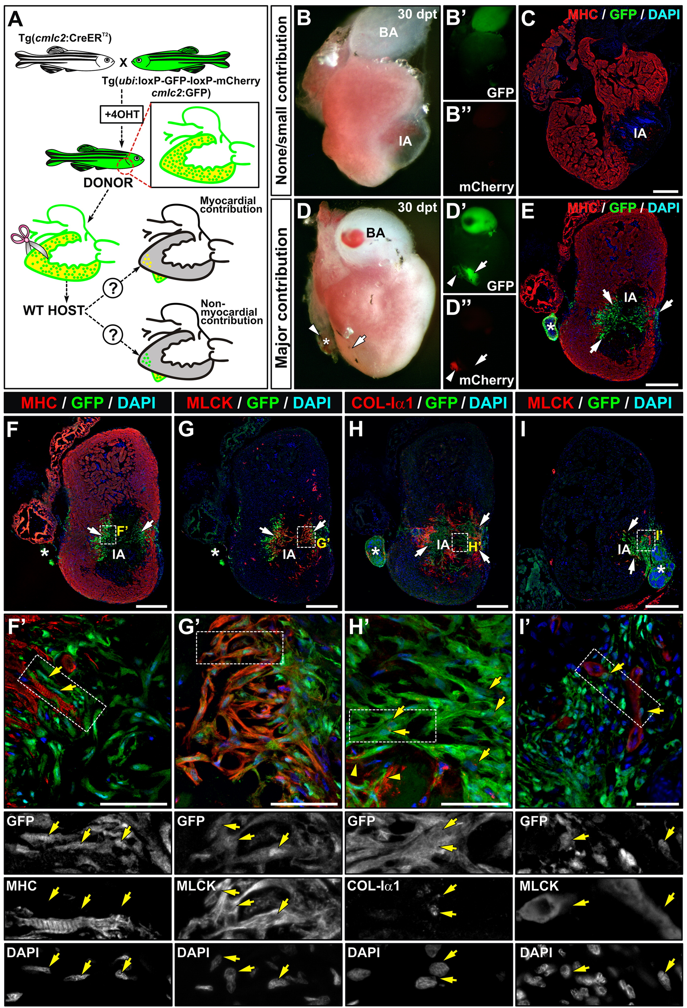
Fig. 5 Long term follow up of transplants. (A) Scheme of the experimental procedure and possible outcomes of transplantation assays using the Tg(cmlc2:CreERT2)/(ubi:loxP-GFP-loxP-mCherry)/(cmlc2:GFP) line as donor. Recombination was induced at larval stages to induce mCherry expression in cardiomyocytes. Grafts of transgenic fish, expressing mCherry and GFP in the myocardium and GFP only in all other cells, were transplanted onto cryoinjured wildtype hearts. If grafted myocardium contributes to the regenerating myocardium, GFP/DsRed cells will be found in the host heart. If any non-myocardial donor cell contributes to the regenerated host myocardium, GFP+ cardiomyocytes would be found in the host heart. (B-B′′) Freshly dissected heart revealing non-detectable graft at 30 dpt, revealed by the absence of signals for GFP (B′) and mCherry (B′′). Note the presence of a large IA and the absence of regeneration. (C) Immunohistochemistry on a section of the same heart as in B. Antibodies used are indicated in the panel. Note that no GFP+ (graft-derived) cells can be found inside the host heart. (D-D′′) Freshly dissected heart revealing no signs of injury at 30 dpt and a visible graft: the host heart contains graft-derived cells (D′) but no graft-derived cardiomyocytes (D′′). (E) Immunohistochemistry on a section of the same heart as in D. Antibodies used are indicated in the panel. Many GFP+ cells are detected inside the host and accumulate at the borders of the remnant IA (arrows). Note the partial regeneration of the myocardial wall. Arrowheads mark graft, arrows mark graft-derived GFP-positive cells. (F-I′) Immunohistochemistry on sections with antibodies indicated above the panels; panels F′-I′ show zoomed views of boxed areas. There is no collocalization of GFP with the myocardial marker MHC, but broad overlap with MLCK and Col-1α1. Asterisks mark the graft or its position in a consecutive section. Bars, 200 μm (full views) and 50 μm (magnifications).
Reprinted from Developmental Biology, 370(2), Manuel González-Rosa, J., Peralta, M., and Mercader, N., Pan-epicardial lineage tracing reveals that epicardium derived cells give rise to myofibroblasts and perivascular cells during zebrafish heart regeneration, 173-186, Copyright (2012) with permission from Elsevier. Full text @ Dev. Biol.