
|
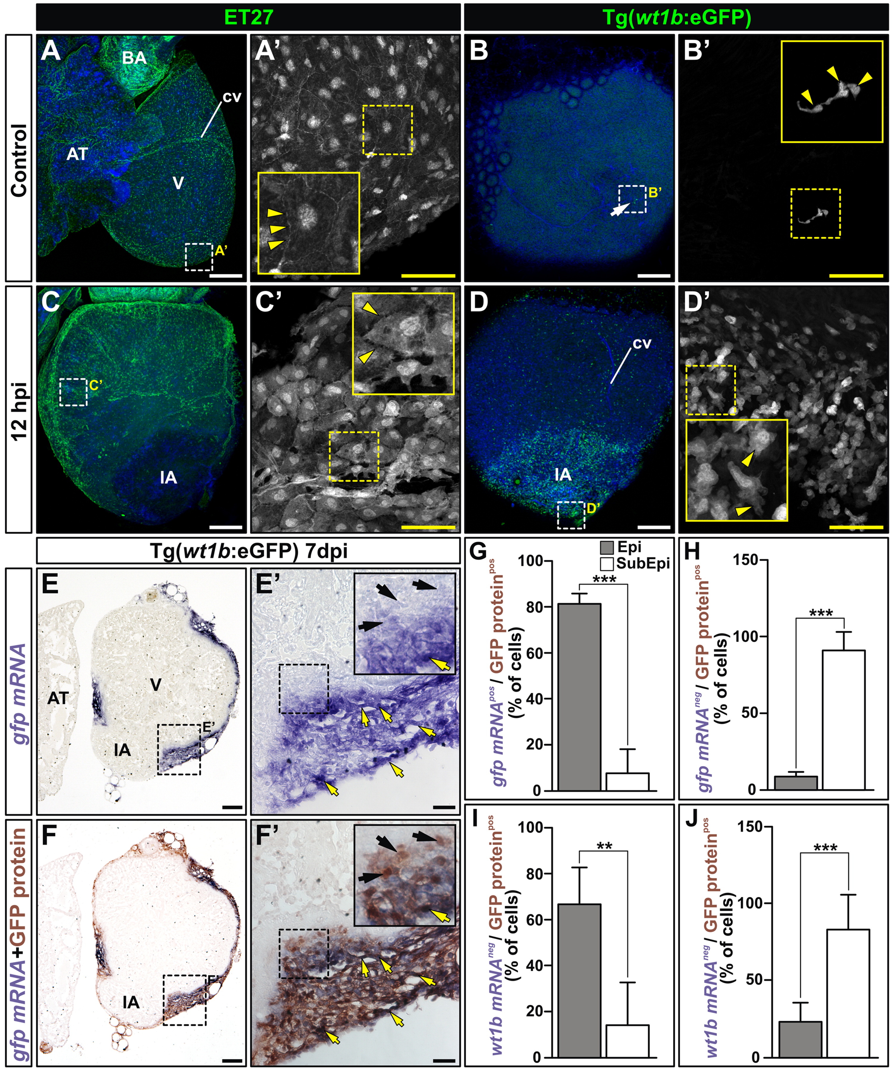
Fig. 1 Epicardial derived cells migrate upon cryoinjury. (A-D) Whole-mount imaging of dissected control and cryoinjured hearts (12 h postinjury: hpi) from the ET27 and Tg(wt1b:eGFP) lines. Nuclei are counterstained with DAPI (blue). A′-D′ are zoomed images showing the GFP channel in the boxed areas in A-D. Arrows marks GFP+ cells, arrowheads mark morphological cell features. (E-J) Comparison of the expression domains of wt1b:GFP protein and gfp and wt1b mRNA. (E,E′) Full view and zoomed images of gfp in situ hybridization on a Tg(wt1b:eGFP) heart section at 7 dpi. Yellow arrows mark cells expressing gfp mRNA, black arrows mark cells devoid of gfp mRNA expression. (F,F′) Same sections in E,E′ after anti-GFP immunohistochemistry. Yellow arrows mark cells coexpressing GFP protein and gfp mRNA, black arrows mark cells only expressing GFP protein. (G) and (H) Percentage of cells coexpressing gfp mRNA and protein (G) or only protein (H). Note that while most epicardial cells coexpress gfp mRNA and protein, most subepicardial cells only express GFP protein. (I) and (J) Percentage of cells coexpressing wt1b mRNA and GFP protein (I) or only protein (J). Results are similar to those in G and H. Data are means of cell percentages±S.D. counted on 1-3 heart sections per specimen from 2-5 specimens (***P<0.001; **P<0.01; one-way ANOVA followed by Tukey′s honest significant difference test; 100-150 cells counted per section). AT, atrium; BA, bulbus arteriosus; cv, coronary vessel; dpi, days postinjury; IA, injured area; ISH, in situ mRNA hybridization; V, ventricle. Bars, 200 μm (full views) and 50 μm (magnifications).
Reprinted from Developmental Biology, 370(2), Manuel González-Rosa, J., Peralta, M., and Mercader, N., Pan-epicardial lineage tracing reveals that epicardium derived cells give rise to myofibroblasts and perivascular cells during zebrafish heart regeneration, 173-186, Copyright (2012) with permission from Elsevier. Full text @ Dev. Biol.