
Fig. 4
- ID
- ZDB-IMAGE-121130-9
- Publication
- Hailey et al., 2012 - Loss of Slc4a1b Chloride/Bicarbonate Exchanger Function Protects Mechanosensory Hair Cells from Aminoglycoside Damage in the Zebrafish Mutant persephone
- All Figures
- Figures for Hailey et al., 2012
|
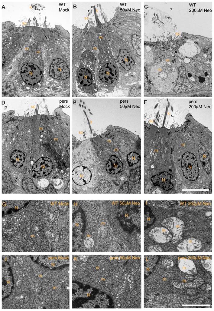
Fig. 4 persephone hair cells have normal morphology and show only modest ultrastructural changes when treated with neomycin.
Zebrafish (5 dpf) treated with or without neomycin were euthanized, tails were removed for genotyping, and heads were fixed for transmission electron microscopy. Images of transverse sections through neuromasts are shown. (N: hair cell nuclei, m:mitochondria, sc: support cell, k: kinocilia. Top panels A–C and bottom panels G–I show hair cells from wildtype siblings. Top panels D–F and bottom panels J–L show hair cells from persephone homozygotes. Panels were either mock-treated or exposed to 50 µM or 200 µM neomycin. Hair cells of mock-treated wildtype siblings and persephone mutants show organized stereocilia, a large central nucleus, and normal mitochondrial morphology. There are no readily apparent differences between the wildtype siblings and persephone mutants. Hair cells of persephone mutants treated with 50 μM neomycin show either mild mitochondrial swelling (E,K) or are indistinguishable from mock-treated wildtype siblings. In comparison wildtype siblings (B,H) show swollen mitochondria and fused stereocilia. Most neuromasts from wildtype siblings treated with 200 µM neomycin lack hair cells, and remaining hair cells exhibit severe damage including condensed nuclei and severely swollen mitochondria or signs of cytolysis (C,I). Hair cells of persephone mutants treated with 200 μM neomycin show minimal damage. Stereocilia are typically oraganized and intact; some but not all hair cells show modestly swollen mitochondria (F,L). See Table 1 for quantification of neomycin-induced damage across genotypes. Top scale bar = 5 μm; Bottom scale bar = 1 μm.