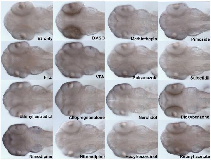
Fig. S1 Analysis of apoptosis in the CNS of drug-treated embryos using TUNEL. Anticonvulsant compounds were administered to 50 hpf embryos for 90 minutes in multi-well plates, at a final concentration of 10 μM. PTZ was administered at a final concentration of 20 mM and VPA was administered at a final concentration of 1 mM. Embryos were fixed and analysed for the presence of apoptotic cells using TUNEL. Each panels shows a dorsal view of the brain of an embryo exposed to the indicated compound. No significant induction of apoptosis was observed after treatment of embryos with any of the selected compounds.
Image
Figure Caption
Acknowledgments
This image is the copyrighted work of the attributed author or publisher, and
ZFIN has permission only to display this image to its users.
Additional permissions should be obtained from the applicable author or publisher of the image.
Full text @ Dis. Model. Mech.

