
Fig. 2
- ID
- ZDB-IMAGE-121116-25
- Genes
- Antibodies
- Publication
- Tessadori et al., 2012 - Identification and functional characterization of cardiac pacemaker cells in zebrafish
- All Figures
- Figures for Tessadori et al., 2012
|
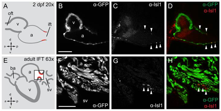
Fig. 2 Isl1 expression in the embryonic and adult zebrafish heart.
Single confocal scans of a fluorescent antibody labeling of Isl1 and eGFP in embryonic (2 dpf) (A–D) and adult (E–H) zebrafish expressing Tg(myl7:eGFP) in all cardiomyocytes. GFP+ cardiomyocytes are displayed in grey (B, F) and in green in (D, H). Isl1 is shown in grey (C, G) and in red (D, H). Arrowheads indicate Isl+/GFP+ cells. Illustrations of a lateral view of a 2 dpf (A) and adult (E) zebrafish heart indicate the location of Isl1+ cells (red). The box in panel E represents the area shown in (F–H). (B–D) Fluorescent immunolabeling of Isl1 and eGFP in a 2 dpf embryo (sagittal section 100 μm). At this time point Isl1+/GFP+ cells were only found in the IFT of the heart. (F–H) Fluorescent immunolabeling of Isl1 and eGFP in an adult zebrafish heart (sagittal section 100 μm). Isl1+/GFP+ cells are located at the junction of the sinus venosus and atrium in the inflow region of the heart (arrowheads). Isl1+ cells showed low expression of myl7. v, ventricle; a, atrium; oft, outflow tract; ift, inflow tract; ba, bulbus arteriosus; sv, sinus venosus; a, anterior; p, posterior; d, dorsal; v, ventral. Scale bars represent 50 μm.