
|
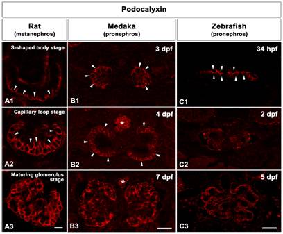
Fig. 4 The progression of podocyte differentiation visualized by Podocalyxin immunostaining.
A1-A3: Cross sections of rat metanephric glomeruli. Throughout the rat metanephric glomeruli development, Podocalyxin is mainly localized at the apical membrane of podocytes. At the S-shaped body stage, Podocalyxin marks the apical membrane of the individual podocytes in a cap-shaped pattern (arrowheads in A1). By early capillary loop stage, Podocalyxin localizes in a U-shaped pattern in the individual podocytes (arrowheads A2). By the maturing glomerulus stage, Podocalyxin staining is detected along the entire surface of podocytes (A3). B1-B3: Cross sections of medaka pronephric glomeruli. Podocalyxin immunostaining is detected in the individual podocytes as a U-shaped pattern at 3 and 4 dpf (B1, B2). At 7 dpf, Podocalyxin staining is detected along the entire surface of podocytes (B3). Asterisks indicate blood cells in dorsal aorta. C1-C3: Cross sections of zebrafish pronephric glomeruli. At 34 hpf, Podocalyxin marks the apical membrane of individual podocytes at 34 hpf (C1). By 2 dpf (C2) and 5 dpf (C3), Podocalyxin immunostaining is expressed along entire surface of podocytes. Scale bars = 10 μm.