
|
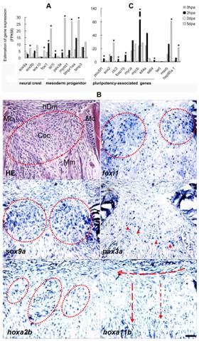
Fig. 6 Genetic identification of blastemal cells.
Transcriptomal analyses of selected cell identity genes at discrete four time points during regeneration (A, C). The marker genes were selected according to the published data (referenced in the text). Figure A shows differential expression of tissue specific progenitor markers of neural crest cell and mesodermal progenitors. isl1 was temporally and highly activated at 2 hpa. foxi1 was activated at 2 hpa and maintained activation at 2 dpa and 5 dpa. The similar expression pattern occurred to pax3a. In contrast, sox9a was not activated until 5 dpa. Figure C shows differential expression of pluripotency-associated genes. It also includes the other three genes (tert, hsp90a1 and msxb), which have been reported to be regeneration-associated. Pluripotency-associated genes were selectively activated at 2 hpa except that sox2 was activated at 5 dpa. Asterisk indicates significant (P<0.05) upregulation of gene expression compared to the previous time point. Figure B shows localization of the cell identity markers in blastema at 5 dpa. The expression domains of foxi1, sox9a and hoxa2b were partially overlapped in the blastema chondrogenesis zone (red ring). hoxa2b was widely expressed with island-like distribution in chondrogenic blastema. pax3a was expressed in the regenerating muscle (arrow head). hoxa11b was highly expressed in blastemal mesenchyme and arranged in the directions toward the hypodermis (horizon arrow) and the mandibular muscle (vertical arrow). The consecutive sections were used for HE staining and RNA-in situ hybridization. Mc, Meckel cartilage; Coc, chondrogenic center; Mm, mandibular muscle; hDm, hypodermal mesenchyme. Scale bars, 50 μm.