
|
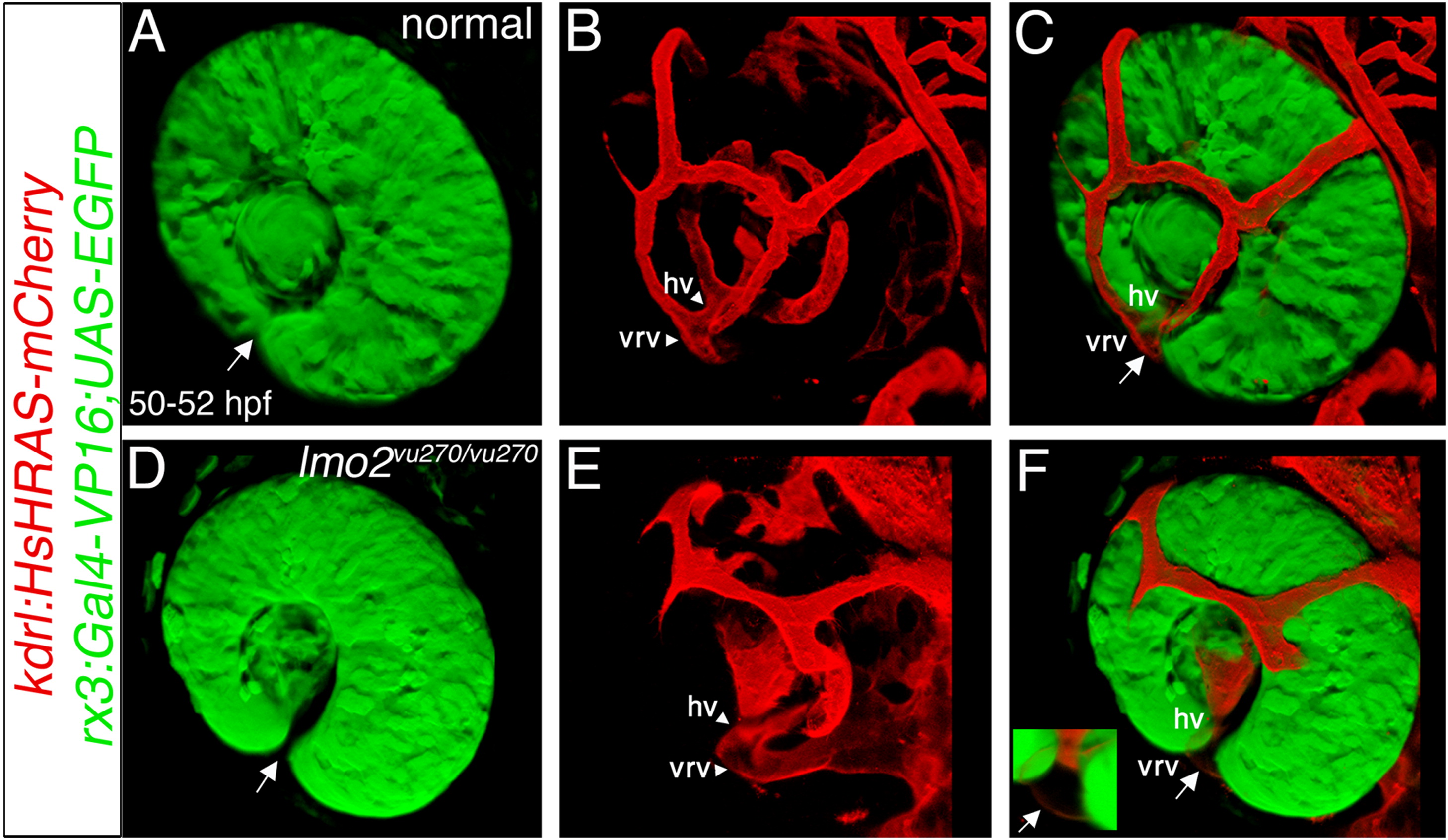
Fig. 5 Dilated vessels are located in the optic fissure. (A–F) Three-dimensional shadow rendering of confocal z-stacks of eyes (green) and ocular vessels (red) in 2 dpf (rx3:Gal4-VP16);(UAS:EGFP);(kdrl:HsHRAS-mCherry) transgenic normal (A–C) and lmo2vu270/vu270 (D–F) embryos. In the merged panels (C and F) most of the hyaloid vessels cannot be seen. (E and F) Growth of superficial ring vessel is delayed in the mutant (compare to B and C). Arrows point at the optic fissure. Inset in F is a higher magnification of the optic fissure area with the arrow pointing at the dilated vessel in the fissure. Vessel abbreviations are as in Fig. 2. All panels are dorsolateral view, anterior to the left.
Reprinted from Developmental Biology, 369(2), Weiss, O., Kaufman, R., Michaeli, N., and Inbal, A., Abnormal vasculature interferes with optic fissure closure in lmo2 mutant zebrafish embryos, 191-198, Copyright (2012) with permission from Elsevier. Full text @ Dev. Biol.