
|
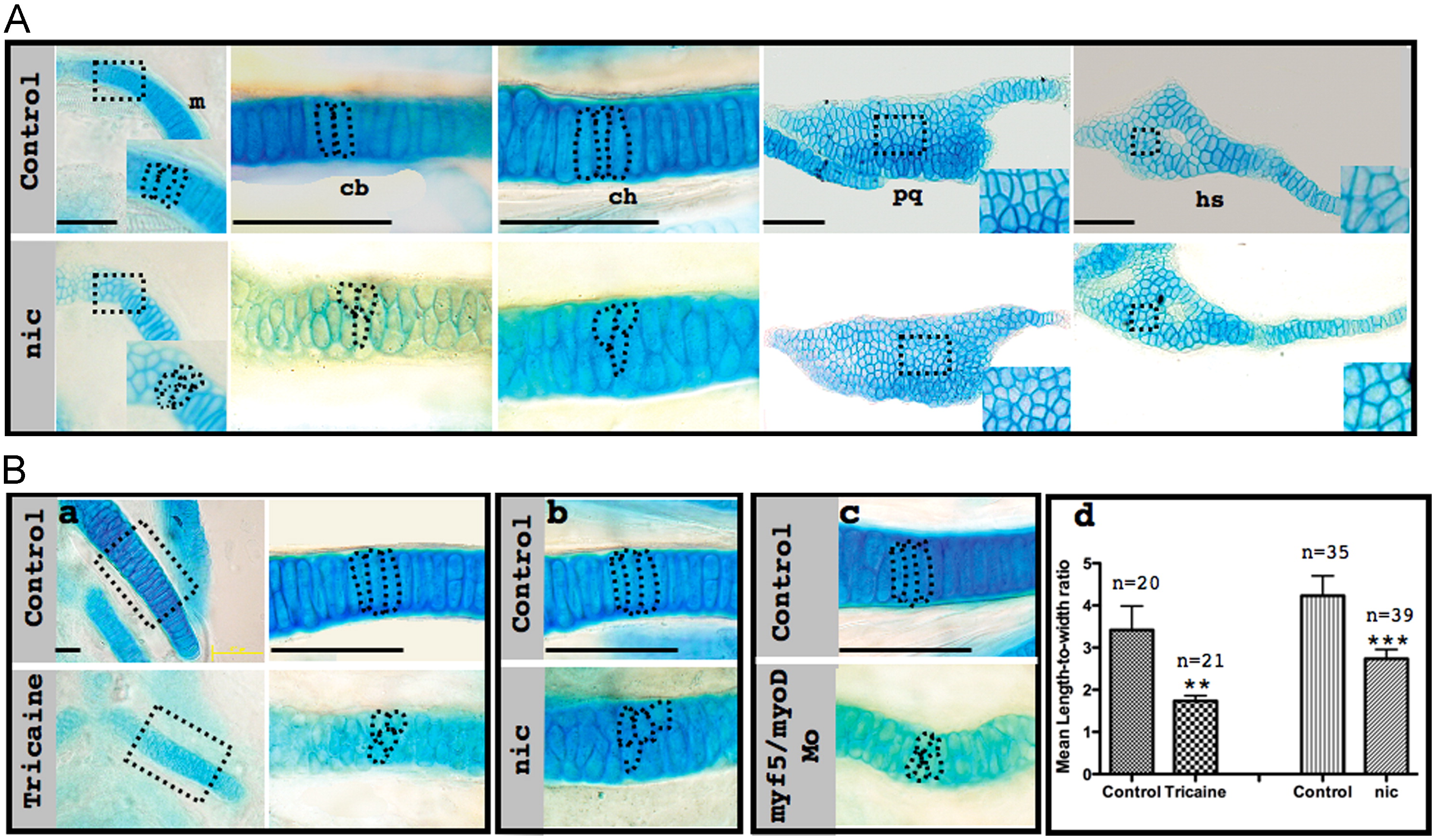
Fig. 3 Muscle contraction is essential for creating proper stacking pattern. (A) Flat-mounted Alcian blue stained control (upper panel) and nic mutant (lower panel) 120 hpf zebrafish show the difference in cell morphology in various skeletal elements. Magnifications of squared areas are shown in the lower right corner. Dashed lines demarcate individual cells. (B) Flat-mounted Alcian blue stained control (upper panel) and tricaine-paralyzed (lower panel) ceratohyal cartilage at 120 hpf (a). Right: Magnification of squared areas; dashed lines demarcate individual cells. Flat-mounted Alcian blue stained ceratohyal cartilage from a corresponding location as shown in (a) from control embryos (upper panels), nic mutant (b, lower panel) and myf5/myod double morphants (c, lower panel) at 120 hpf. Quantification of length-to-width ratio (d) in cells from the middle part of the ceratohyal cartilage of control, tricaine-paralyzed (F13.887=35.63, p=0.004) and nic mutant (F1,72=55.44, p=1.65E-10) embryos; n represents number of measured cells; scale bars represent 50 μm.
Reprinted from Developmental Biology, 370(1), Shwartz, Y., Farkas, Z., Stern, T., Aszódi, A., and Zelzer, E., Muscle contraction controls skeletal morphogenesis through regulation of chondrocyte convergent extension, 154-163, Copyright (2012) with permission from Elsevier. Full text @ Dev. Biol.