
|
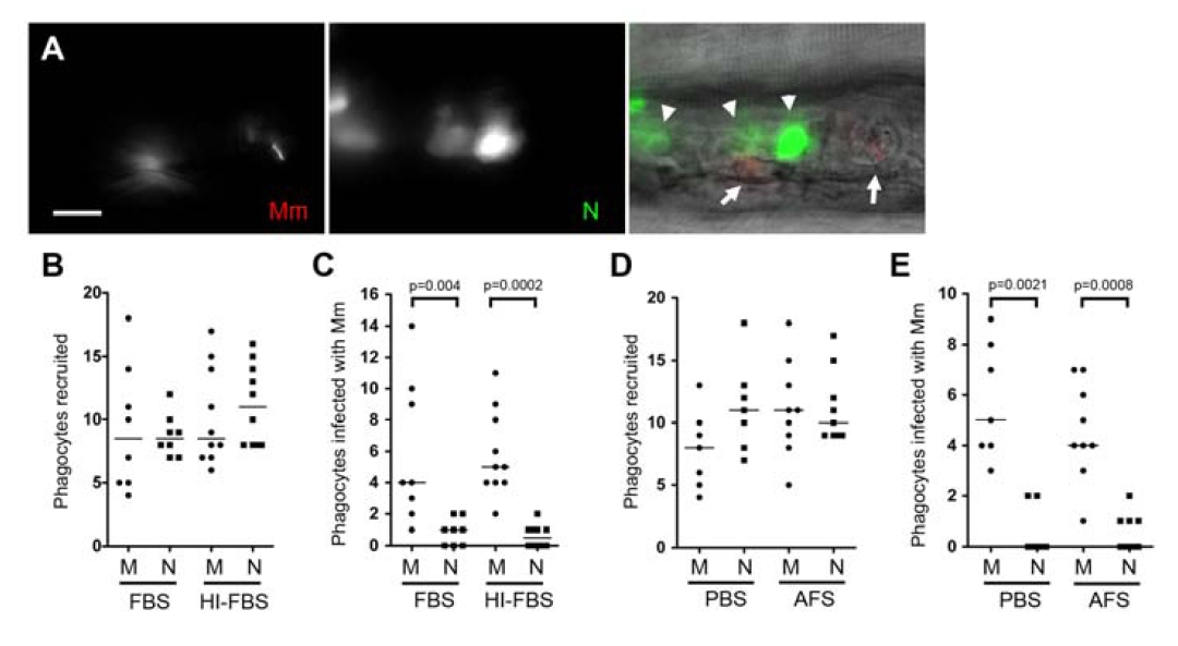
Fig. S1
(Related to Figure 2) Mycobacteria are poorly phagocytosed by neutrophils at the initial infection site at late developmental stages or in the presence of serum
(A) Fluorescence and DIC overlay of red M. marinum (Mm) (arrows) and green neutrophils (N) (arrowhead) in 5 dpf larva 24 hours after caudal vein injection. Scale bar, 10 μm. (B and C) Median phagocyte recruitment and bacterial phagocytosis six hours after HBV co-injection of 80-120 M. marinum and 30-40 P. aeruginosa, both incubated with fetal bovine serum (FBS) or heat-inactivated (HI) FBS. (D and E) Median phagocyte recruitment and bacterial phagocytosis phagocytosis six hours after HBV co-injection of 90 M. marinum, and 30-40 P. aeruginosa incubated with adult fish serum (AFS) or phosphate buffered saline (PBS). M, macrophage; N, neutrophil. p values by Mann- Whitney rank test.