
Image
|
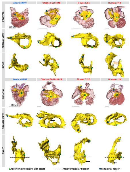
Figure Caption
Fig. 5
Three-dimensional reconstructions of Tbx3 (yellow) expression in amniotes reveal a shared design.
Reconstructions are based on in-situ hybridizations of serial sections, except in human (based on immunohistochemistry, modified from [57], [58]). The Tbx3 domains are strikingly similar in the early phases of chamber formation (upper panel). The Tbx3 expression of the Anolis ventricle is very similar to that associated with ventricular septation (black arrows) in the other amniotes (lower panel).
Acknowledgments
This image is the copyrighted work of the attributed author or publisher, and
ZFIN has permission only to display this image to its users.
Additional permissions should be obtained from the applicable author or publisher of the image.
Full text @ PLoS One