
|
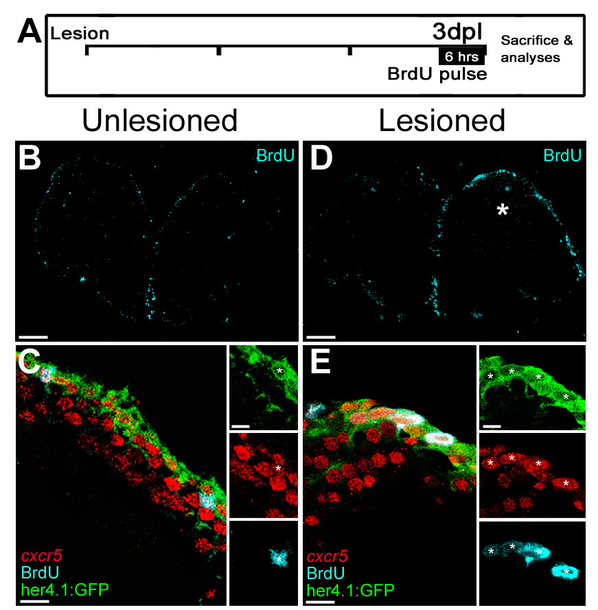
Fig. 2 cxcr5 is expressed in proliferating radial glial cells (RGCs). (A) Scheme for experimental setup. At 3 days after a lesion or a sham operation, bromo-deoxyuridine (BrdU) is applied for 6 hours before sacrificing the animals. (B) BrdU immunohistochemistry on the unlesioned (sham-operated) adult zebrafish telencephalon section showing the proliferating cells. (C)cxcr5 fluorescent in situ hybridization (FISH) coupled to BrdU and GFP immunohistochemistry on unlesioned Tg(her4.1:GFP) transgenics. Insets show single channel images. BrdU-positive RGCs express cxcr5 (white asterisk). (D) BrdU immunohistochemistry on the 3 days post-lesion (dpl) adult zebrafish telencephalon section. Asterisk indicates the lesion site. (E)cxcr5 FISH coupled to BrdU and GFP immunohistochemistry on 3 dpl Tg(her4.1:GFP) transgenics. Insets show single channel images. BrdU-positive RGCs increase in number and they express cxcr5 (white asterisks). Scale bars 50 μm in B and D, and 10 μm in C and E; n = 3 telencephalons.