
|
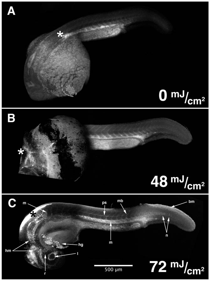
Fig. 4 HxBP can be UV-photocrosslinked in vivo, and detected in situ by confocal microscopy.
Composite confocal micrographs of 24 hpf zebrafish embryos injected anteriorly with 50 μM trifunctional HxBP probe, recovered, and exposed to increasing levels of UV irradiation reveals increasingly spatially structured patterns of fluorescence up to 72 mJ/cm2. Structures exhibiting strong HxBP labeling in 24 hpf embryos include the retina (r) and lens (l), head mesenchyme (hm), hatching gland (hg), perichordal sheath (ps), isolated notochord cells in the elongating region of the notochord (n), maturing myotome boundaries (mb), mesenchymal tissues of the trunk and tail (m), and the basement membrane underlying the epithelium dorsal to the elongating tail (bm). Asterisks mark the point of injection.