
|
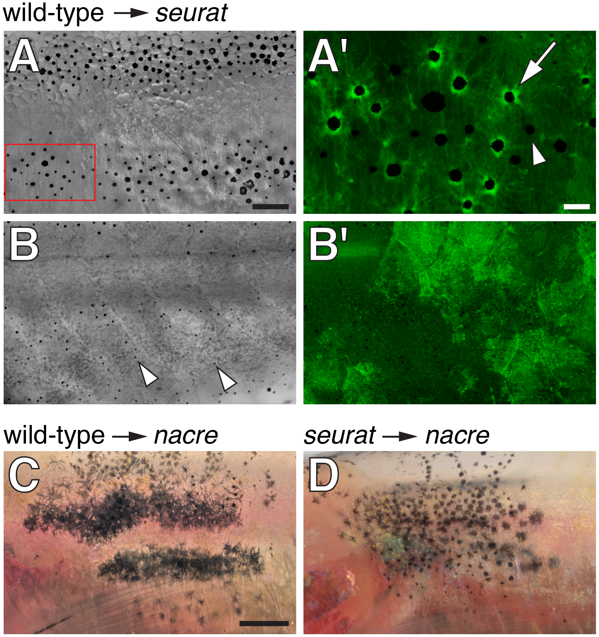
Fig. 2
seurat is required autonomously to the melanophore lineage.
(A, B) Wild-type Tg(bactin:GFP) cells transplanted to seurat mutant hosts. Fish shown are juveniles (~13 mm standardized standard length, SSL [11]) and were treated just prior to imaging with epinephrine, which contracts melanosomes towards cell bodies, thereby facilitating the detection of GFP fluorescence. (A) Chimeras that developed wild-type melanophores exhibited patches of restored stripes (n = 6). (A2) Detail of boxed region in A, showing GFP+ melanophores (e.g., arrow), as well as occasional GFP, presumptive seurat mutant melanophores (e.g., arrowhead). (B) Chimeras in which wild-type melanophores failed to develop exhibited a seurat mutant pattern of dispersed melanophores (arrowheads; n>100). In the example shown here, wild-type GFP+ cells developed as epidermis (B2 shown at same magnification as B). (C) When wild-type melanophores differentiated in a nacre mutant background, patches of normal stripes developed (n = 3; [16]). (D) By contrast, when seurat mutant melanophores differentiated in nacre hosts, these cells retained a dispersed pattern, as in the seurat mutant (n = 8), indicating a failure of xanthophores, iridophores, or other cell types to rescue melanophore stripe organization. In additional experiments, in which nacre; Tg(bactin:GFP) cells were transplanted to seurat mutant hosts, the differentiation of nacre- GFP+ (seurat+) iridophores likewise failed to rescue melanophore stripes in the seurat mutant background (donor xanthophores did not develop in these chimeras; data not shown). Scale bars: in (A) 100 μm for (A,B,B2); in (A2) 20 μm for (A2); in (C) 500 μm for (C,D).