
Fig. 4
- ID
- ZDB-IMAGE-120830-5
- Publication
- Yan et al., 2012 - Slc39a7/zip7 Plays a Critical Role in Development and Zinc Homeostasis in Zebrafish
- All Figures
- Figures for Yan et al., 2012
|
Fig. 4
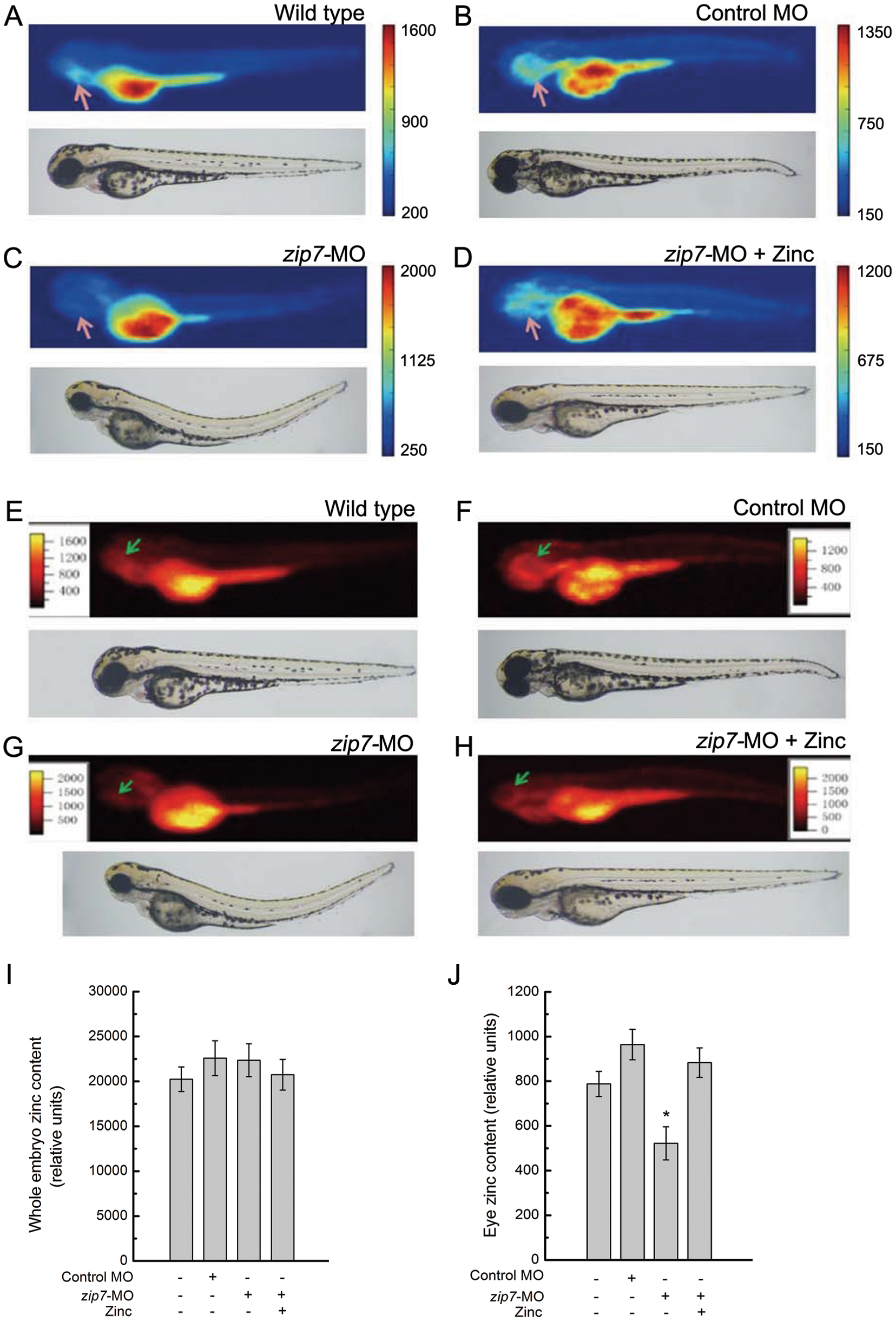
SR-XRF images and quantitative analysis of Zinc in zip7-Deficient Embryos and Zn2+ (50 μM) rescue.
Lateral views (anterior to the left) of embryos at 72 hpf, arrows indicate eye. (A and E) Wild type without any microinjection, (B and F) wild type with control-MO microinjection (2 nL, 10 ng), (C and G) Wild type with zip7-MO microinjection (2 nL, 10 ng), (D and H) Wild type with zip7-MO microinjection (2 nL, 10 ng) and hatching in the presence of Zn2+ (50 μM). (A–D) Two-dimensional images of SSRF, (E–H) Three-dimensional images of SSRF. Quantitation and statistical analysis of zinc densities in wild type, control-MO, zip7-MO, and zip7-MO+zinc embryos (I and J). (I) Relative zinc content in the whole embryo, and (J) relative zinc content in the eye. *Statistical differences with corresponding wild type (t test, P<0.05). The zinc relative content was acquired by d4/d2/d1 (d4: photon counting of zinc correspondence, d2: electronic counting of light intensity, and d1: irradiation time).