
Fig. 8
- ID
- ZDB-IMAGE-120815-47
- Genes
- Publication
- Liu et al., 2012 - betaPix plays a dual role in cerebral vascular stability and angiogenesis, and interacts with integrin alphavbeta8
- All Figures
- Figures for Liu et al., 2012
|
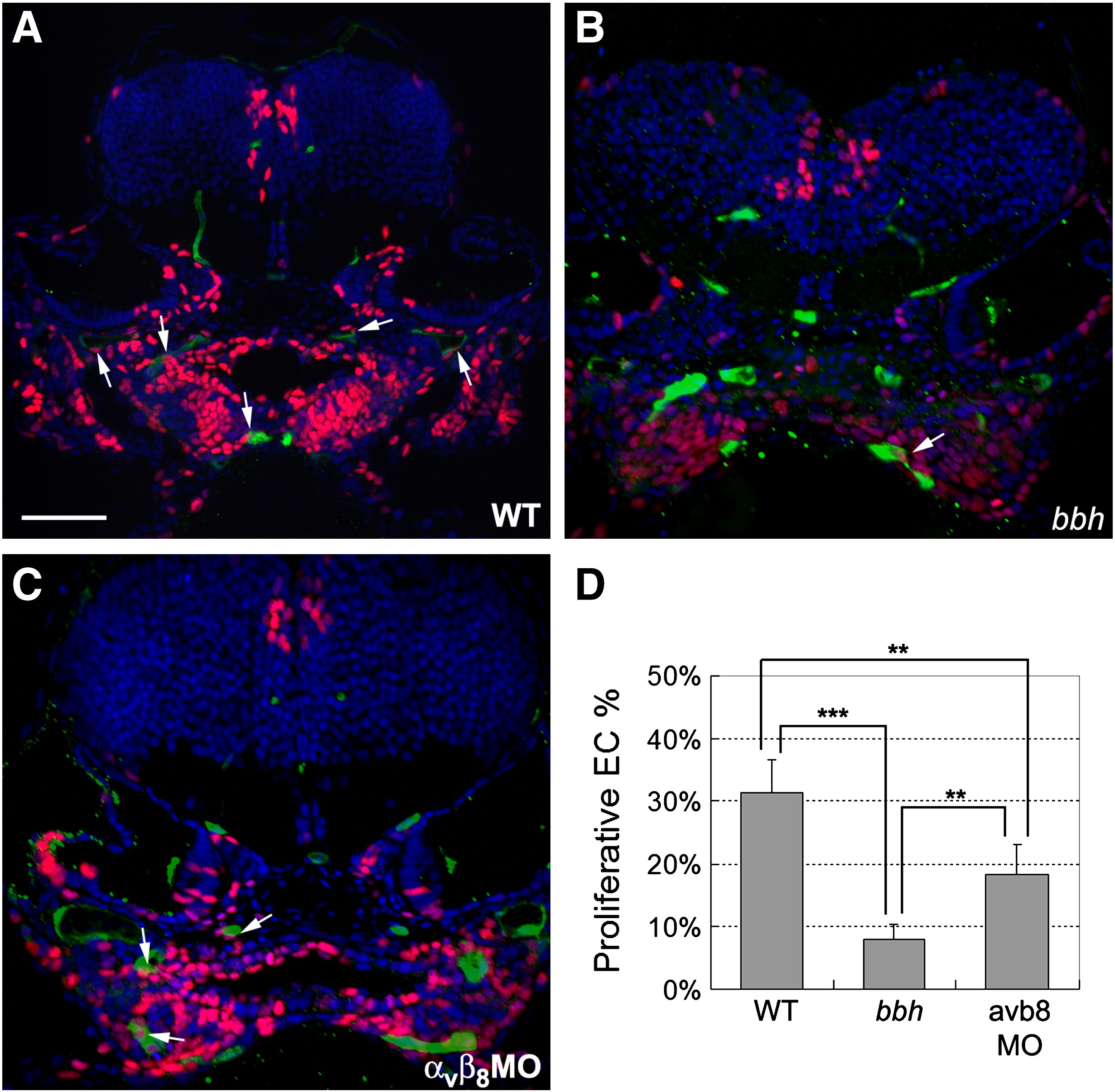
Fig. 8 Defective endothelial cell proliferation in bbh mutants and integrin αvβ8 double morphants. (A–C) Cell proliferation was detected by pulsing with EdU at 48 hpf and fixation at 54 hpf (red), while endothelial cells are marked by expression of GFP in the Tg(kdrl:eGFP)la116 line (green) and nuclei are stained with DAPI (blue). In general, higher levels of EdU incorporation are detected in wild type embryos (A) as compared to bbh homozygotes (B) or and integrin αvβ8 double morphants (C). Arrows highlight proliferative endothelial cells. (D) The percentage of proliferative endothelial cells were analyzed from the percentage of cells with co-staining of EdU and GFP vs. the total number of GFP cells as observed on sections from 5 wild types, 5 bbh homozygotes, and 5 integrin αvβ8 double morphants. Significance was determined by a Student′s t test, where ** represents p < 0.005, and *** represents p < 0.001.
Reprinted from Developmental Biology, 363(1), Liu, J., Zeng, L., Kennedy, R.M., Gruenig, N.M., and Childs, S.J., betaPix plays a dual role in cerebral vascular stability and angiogenesis, and interacts with integrin alphavbeta8, 95-105, Copyright (2012) with permission from Elsevier. Full text @ Dev. Biol.