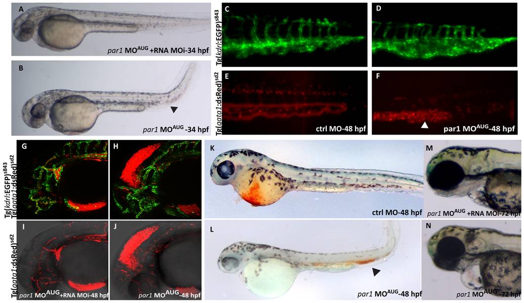
Fig. 3 par1 knockdown causes ICM blood pooling, heart edema and head hemorrhage.
(A) 34 hpf rescued embryo with heart beat and blood flow, resembling a ctrl MO embryo at this stage. (B) par1 morphant showing blood pooling (arrowhead) with bend tail and heart edema. (C–F) The posterior part of the embryo at 48 hpf. The ctrl MO embryo has dorsal aorta, CV, ISVs and arteries with expanded lumen with blood flow as seen in the double transgenic background (C and E). The par1 morphant shows ISVs, arteries, dorsal aorta and CV, but the blood cells are pooled in the CV (arrowhead in F) and the vein is bulged and malformed (asterisk in D). (G–J) Head of 48 hpf double transgenic embryos, displaying endothelial cells in green fluorescence and blood cells in red fluorescence (Tg(kdrl:EGFP)s843; Tg(gata1:dsRed)sd2). The blood vessels in the head of both the rescued embryo and the morphant are developed normally, but there is a leakage of blood cells into the mesencephalon of the morphant embryo, causing hemorrhage. Blood flow in the body of both rescued and morphant embryo was normal (not shown). (K–L) O-dianisidine staining at 48 hpf. (K) embryo treated with Ctrl MO displaying normal blood circulation; (L) embryo treated with par1-MOAUG showing reduced level of Hb in the Duct of Cuvier and blood pooling in the tail. (M–N) Embryos at protruding mouth stage (72 hpf). Contrary to the rescue embryo (M), the par1 morphant has a slight cardiac edema, no mouth protrusion and no visible swim bladder (N).

