
|
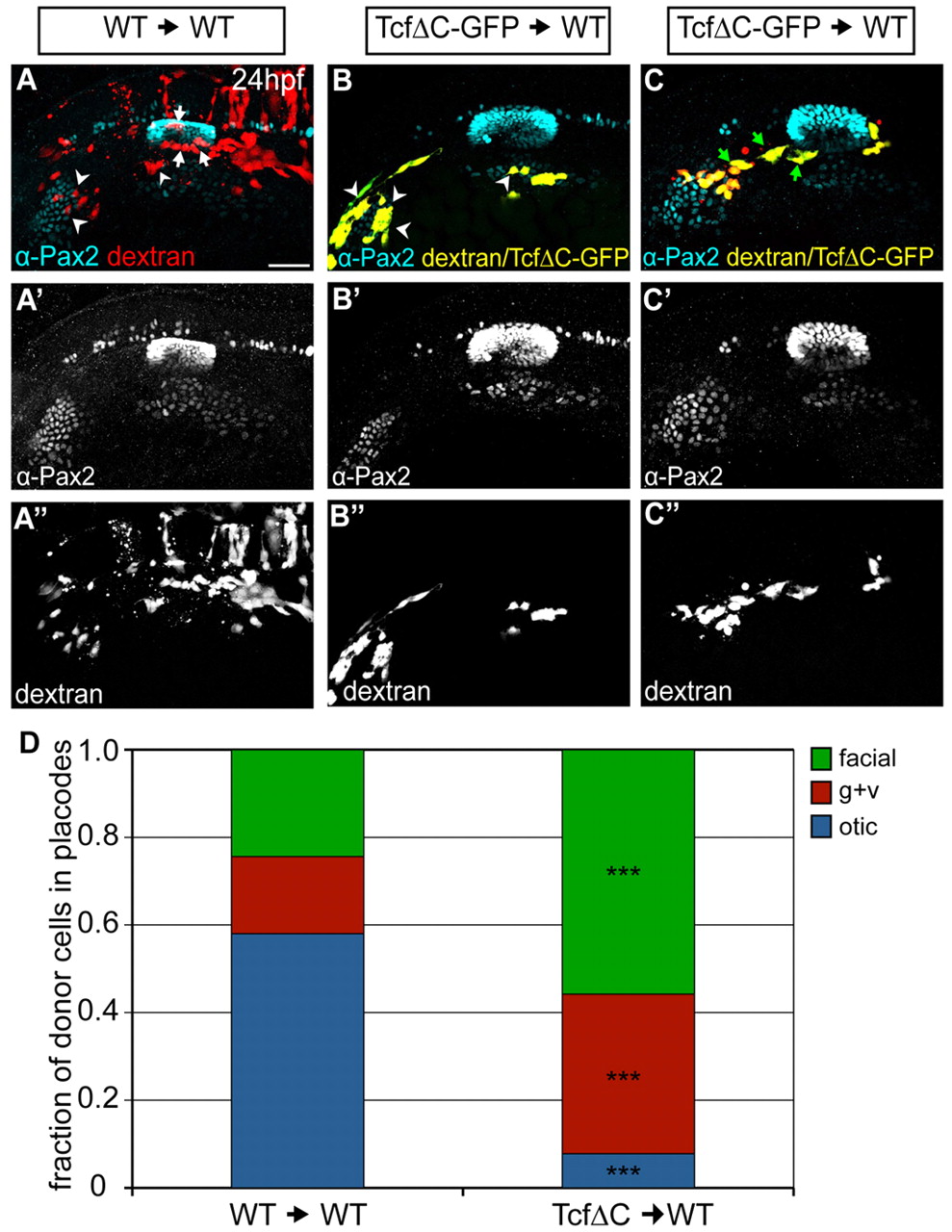
Fig. 8 Wnt activation is required cell-autonomously for otic commitment. (A-C′′) Wild-type zebrafish hosts containing rhodamine-dextran-positive cells from wild-type (red, A) or Tg(hsp70:tcfΔC-EGFP) (yellow, B,C) donors were heat-shocked at 10 hpf and immunolabeled for Pax2a (cyan) at 24 hpf. Cells derived from Tg(hsp70:tcfΔC-EGFP) donors appear yellow owing to colocalization of EGFP and rhodamine-dextran. Arrowheads indicate donor cells in EB placodes; white arrows mark donor cells in the otic vesicle, green arrows mark donor cells in presumptive gAll/gVIII. (D) Relative contributions of donor cells to otic vesicle and EB placodes at 24 hpf in wild-type and Tg(hsp70:tcfΔC-EGFP) transplants. Tg(hsp70:tcfΔC-EGFP)+ cells are biased towards facial and glossopharyngeal/vagal placodes, rarely segregating to the otic vesicle [total of 77 and 131 donor cells counted from 11 Tg(hsp70:tcfΔC-EGFP) and ten wild-type transplants, respectively; χ2-test; ***P<<0.001]. Scale bar: 50 μm.