
|
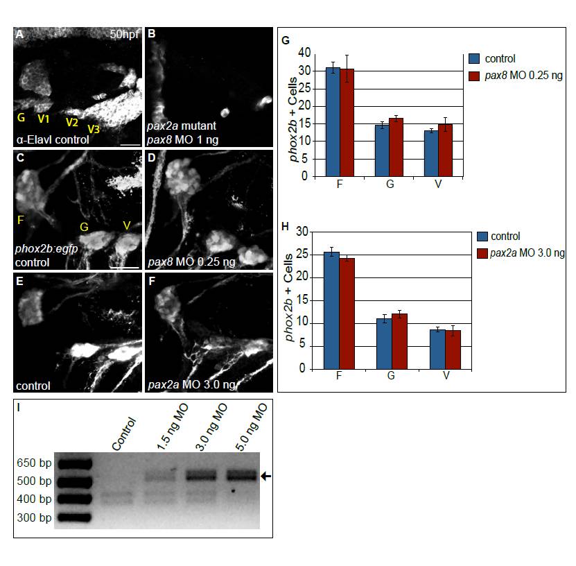
Fig. S5 Combined activity of Pax2a and Pax8 is necessary for formation of the EB ganglia. (A,B) Embryos derived from a pax2ab593/+ incross immunolabeled with anti-Elavl3/4 to mark differentiated EB neurons in control (A) and embryos injected with 1 ng of pax8-MO (B). Note near-complete loss of Elavl3/4+ neurons in the EB ganglia of the pax2a mutant that also received pax8-MO (B). (C-F) Confocal projection from 50 hpf Tg(phox2b:EGFP)w37 embryos. Control embryos (C,E) were compared with embryos injected with either 0.25 ng of pax8-MO (D) or 3.0 ng of pax2a-MO (F). (G,H) Quantification of phox2b+ neurons at 50 hpf in the facial, glossopharyngeal and most anterior small vagal ganglia in embryos injected with 0.25 ng of pax8-MO (G) and 3.0 ng pax2a-MO (H) versus controls (n≥34 cells from five embryos per condition). Note the lack of significant difference in ganglion sizes when MOs are singly injected at suboptimal concentrations. Error bars represent s.e.m. (I) RT-PCR of pax2a splice variant products from embryos that were injected with increasing amounts of pax2a MO. The amount of an RT-PCR product corresponding to an aberrant splice variant (arrow) increased with respect to injected pax2a-MO in a dose-dependent manner. Scale bars: 25 μm.