
Image
|
Figure Caption
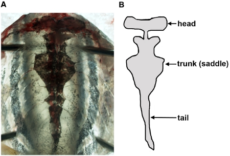
Fig. 2
Visualization of the zebrafish kidney in an unfixed sample. Following removal of organs in the body cavity, the kidney appears as a single, flattened organ that is adherent to the dorsal body wall via connective tissues (A), and has been schematized (B) to show its anatomical shape.
Figure Data
Acknowledgments
This image is the copyrighted work of the attributed author or publisher, and
ZFIN has permission only to display this image to its users.
Additional permissions should be obtained from the applicable author or publisher of the image.
Full text @ J. Vis. Exp.