
IMAGE
Fig. S5
- ID
- ZDB-IMAGE-120720-56
- Publication
- Clark et al., 2012 - Loss of Llgl1 in retinal neuroepithelia reveals links between apical domain size, Notch activity and neurogenesis
- All Figures
- Figures for Clark et al., 2012
Image
|
Figure Caption
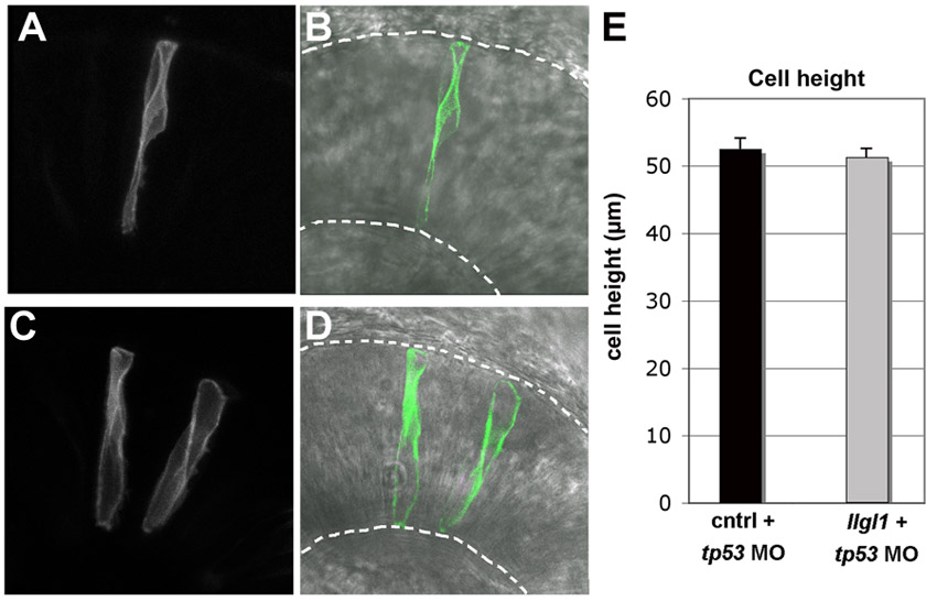
Fig. S5 Cell height of llgl1 morphant retinal neuroepithelia. (A,C) Fluorescent and (B,D) fluorescent with transmitted light images of either (A,B) wild-type donor cells or (C,D) llgl1+tp53 morphant donor cells. Donor cells carried a membrane associated mGFP. Broken lines show apical (top) and basal (bottom) boundaries. (E) Average cell heights of control and llgl1+tp53 morphant cells. For each condition, n=20 cells from more than 5 embryos. No significant difference between groups; P=0.59, Students t-test.
Acknowledgments
This image is the copyrighted work of the attributed author or publisher, and
ZFIN has permission only to display this image to its users.
Additional permissions should be obtained from the applicable author or publisher of the image.
Full text @ Development