
Image
|
Figure Caption
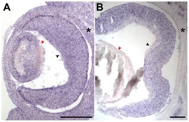
Fig. 6 Tpo is specifically expressed in developing mouse retinas.
In situ hybridization was conducted with a riboprobe specific for Tpo on 20-μm-thick cryosections obtained from mouse embryos at E13.5 (A; transverse section) and P0 (B; coronal section). (A) At E13.5, there was a general expression of Tpo in the whole embryo. (B) By P0, there was a specific expression of Tpo in the retina (black arrowhead) compared with lens (red arrowhead) and the surrounding periocular tissue (asterisks). Scale bar = 200μm for both (A) and (B).
Acknowledgments
This image is the copyrighted work of the attributed author or publisher, and
ZFIN has permission only to display this image to its users.
Additional permissions should be obtained from the applicable author or publisher of the image.
Full text @ PLoS One