
|
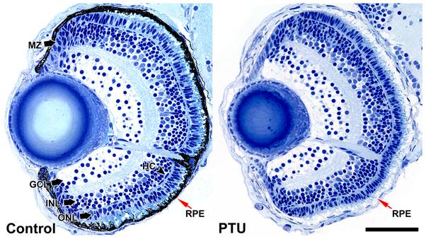
Fig. 2 Histological analysis of zebrafish eyes before and after PTU treatment.
Histological analysis was conducted on 1-μm-thick plastic sections of PTU-treated and control larvae at 4 dpf. The number of different recognizable cell types and categories was counted and the area of the retinas measured. Five sections were analyzed per condition. There are no differences in the counts for all cell types and categories, but the dorsal retinal and lens areas are smaller while the whole retinal area and cell density are marginally higher in the PTU-treated eyes. MZ: marginal zone, HC: horizontal cells, GCL: ganglion cell layer, INL: inner nuclear layer, ONL: outer nuclear layer. The RPE is indicated by red arrows. Scale bar = 50 μm.