
Fig. 1
- ID
- ZDB-IMAGE-120710-15
- Publication
- Ferri-Lagneau et al., 2012 - Ginger Stimulates Hematopoiesis via Bmp Pathway in Zebrafish
- All Figures
- Figures for Ferri-Lagneau et al., 2012
|
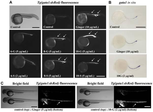
Fig. 1 Ginger extract and its purified phenolic compounds promote Tg(gata1:dsRed) fluorescence and gata1 mRNA expression.
(A) Bright field (top left) and Tg(gata1:dsRed) fluorescence of zebrafish embryos at about 22 hpf, before the onset of circulation (anterior to the left). Exposure to ginger extract or its compounds 8-gingerol (8-G), 10-gingerol (10-G), 8-shogaol (8-S) and 10-shogaol (10-S) promoted Tg(gata1:dsRed) fluorescent erythroid cell development in the ICM and PBI (arrows), as compared to control embryos. N = 35 embryos per group. In this panel, we show an embryo treated with a lower concentration of 6-S (2 μg/ml) as this compound was toxic at higher doses. Scale bar = 400 μm. (B) Whole-mount in situ hybridization of ginger or 10-G treated embryos (8 hpf to 21 hpf exposure) revealed increased expression of gata1 transcript at 22 hpf. N = 50 embryos per group. Scale bar = 350 μm. (C) At 48 hpf, control embryos at the top; ginger or 10-G treated embryos at the bottom. Scale bar = 500 μm. Fluorescent erythrocytes circulating in the axial vasculature (arrows) and in the pericardial space (arrow heads).