
|
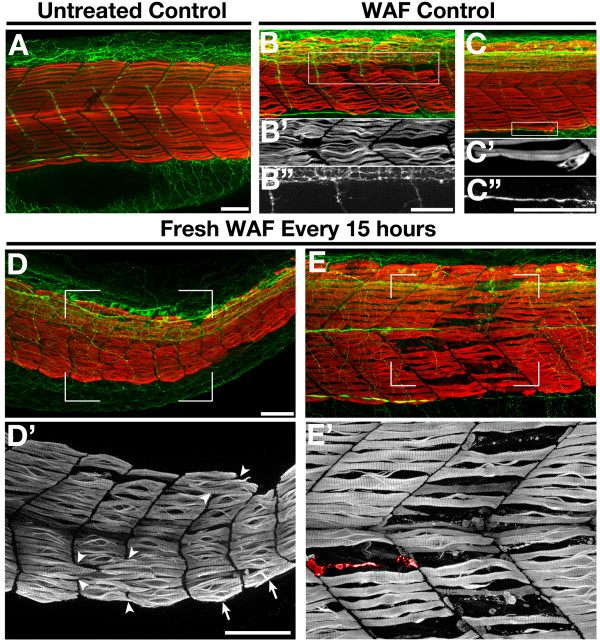
Fig. 9 Repeated application of freshly mixed WAF reproduced the severe skeletal muscle phenotypes. (A-E) Lateral views of the trunk of an untreated control embryo (A), WAF-treated embryos from 3.5 hpf to 48 hpf (B, C), and embryos exposed to freshly mixed WAF every 15 h (D, E). WAF-treated controls showed mild slow muscle defects (B′, C′), while embryos treated repeatedly with refreshed WAF displayed severe somite and slow muscle phenotypes (D′, E′). As seen in earlier experiments, slow muscle fibers spanned presumptive boundaries (D′, arrowheads), somitic shape was irregular (D′, arrows), and slow muscle degeneration was evident (E, a representative degenerating myofibril is pseudo-colored red). Scale bars = 50 μm, A-E′.