
IMAGE
Fig. S5
- ID
- ZDB-IMAGE-120601-72
- Publication
- van der Velden et al., 2012 - The Polycomb group protein Ring1b is essential for pectoral fin development
- All Figures
- Figures for van der Velden et al., 2012
Image
|
Figure Caption
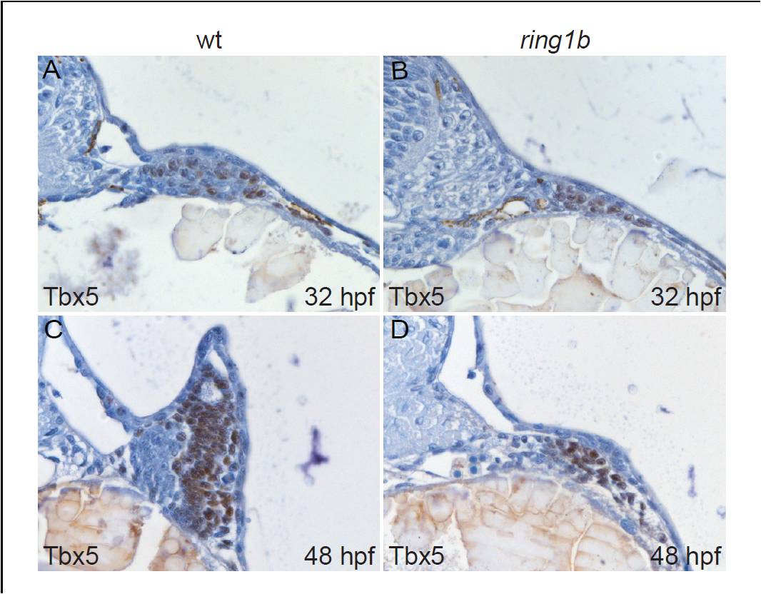
Fig. S5 Nuclear Tbx5 localization in the ring1b pectoral fin mesenchyme. Transverse sections of wild type (A,C) and ring1b mutants (B,D) stained with an antibody against Tbx5 at 32 (A,B) and 48 hpf (C,D). Nuclei in the pectoral fin mesenchyme of ring1b mutants stain positive for Tbx5 at 32 and 48 hpf (B,D).
Acknowledgments
This image is the copyrighted work of the attributed author or publisher, and
ZFIN has permission only to display this image to its users.
Additional permissions should be obtained from the applicable author or publisher of the image.
Full text @ Development