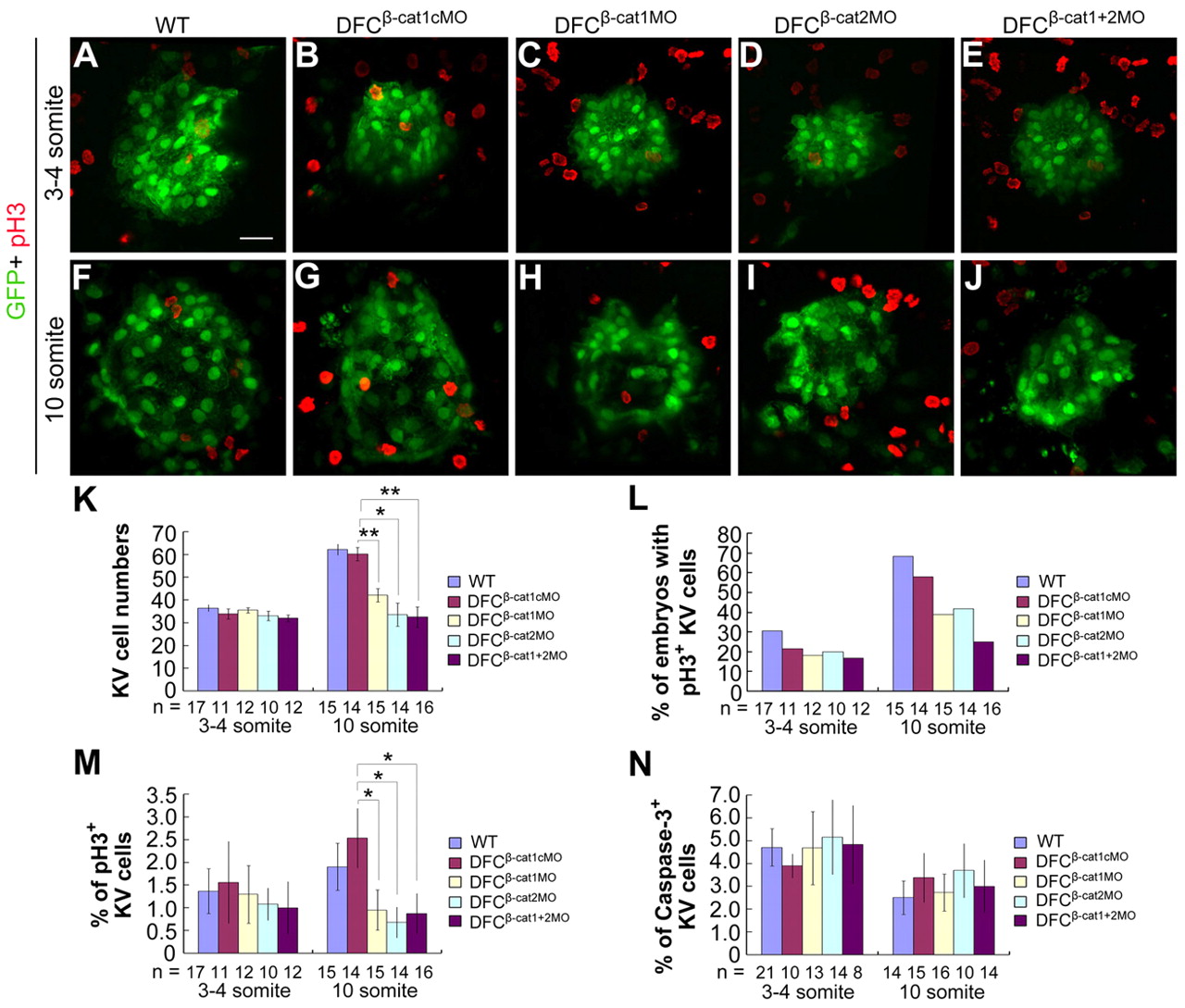
Fig. 7
DFCs knockdown of ctnnb1 and ctnnb2 inhibits KV cell proliferation. Tg(sox17:GFP)s870 transgenic embryos were injected with individual or mixed morpholinos at the 512-cell stage and analyzed later, as indicated. (A-J) Confocal images of KVs at the three- to four-somite (A-E) and 10-somite stages (F-J) following immunostaining with anti-GFP and anti-pH3 antibodies. (K) KV (GFP-positive) cell number was counted at different stages. (L) The percentage of embryos with pH3-positive KV cells. (M) The average percentage of pH3-positive KV cells per embryo. The percentage was calculated for each embryo and then averaged among all observed embryos. The same batch of embryos was subjected to different analyses shown in K-M. (N) The average percentage of caspase 3-positive KV cells per embryo. The injected embryos were co-immunostained with anti-GFP and anti-active caspase 3 antibodies at different stages and observed by confocal microscopy. *P<0.05; **P<0.01; Error bars indicate ±s.e.m.; n, analyzed embryo number.

